Ventricular rhythms
Sinus Rhythm Converting to Ventricular Tachycardia on an EKG Strip
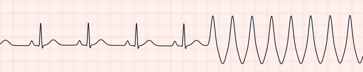
Sinus rhythm converting to ventricular tachycardia is a strip that catches the change as it happens. The first seconds show ordinary sinus beats: a P wave, a narrow QRS, a rate between 60 and 100. Then a wide beat arrives ahead of schedule and is followed by another and another, fast and evenly spaced, and no P wave leads any of them. When the paper runs out the wide beats are still coming.
Criteria from the American Heart Association guidelines and published ECG standards · Every strip checked by measurement · Sources and method · Updated November 15, 2026
- Rate
- 60–100, then > 100
- PR interval
- 0.12–0.20 s, then none
- QRS
- Narrow, then ≥ 0.12 s
How to identify sinus rhythm converting to V-tach in five steps
Ask the same five questions of every strip. The right-hand column is what each one finds on the strip above.
1Rate
Two rates, and both are written down: the sinus rate before the change, and the rate of the wide beats after it, which is above 100 and often 150 to 200.
68 bpm, then 171 bpm
2Rhythm
Regular, then regular again at a much shorter interval. The change itself takes a single beat.
Regular, then a fast regular run
3P waves
Present ahead of each narrow QRS at the start. None can be picked out once the wide beats begin.
1 per QRS, then none
4PR interval
Within the normal range and unchanging while the sinus beats last. There is nothing to measure in the run.
0.16 s on sinus beats
5QRS
Narrow at first. From the first early beat onward, 0.12 seconds or wider, all of one shape, each running into the next.
0.08 s, then wide
What is happening in the heart
Ventricular tachycardia usually starts with a single premature ventricular beat that arrives at the wrong moment. In a ventricle with scarred or poorly supplied muscle, that impulse can find a path that leads back to where it began and start travelling round it. Each lap sets off the ventricles again, far faster than the sinus node was driving them. The sinus node goes on firing in the background, but its impulses reach ventricles that have just been activated and cannot answer, and its P waves are lost inside the wide complexes. The atria and the ventricles are now beating separately.
More sinus rhythm converting to V-tach strips: no two are the same
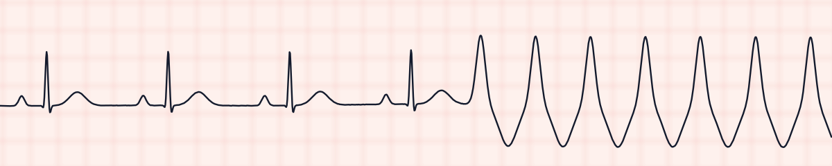
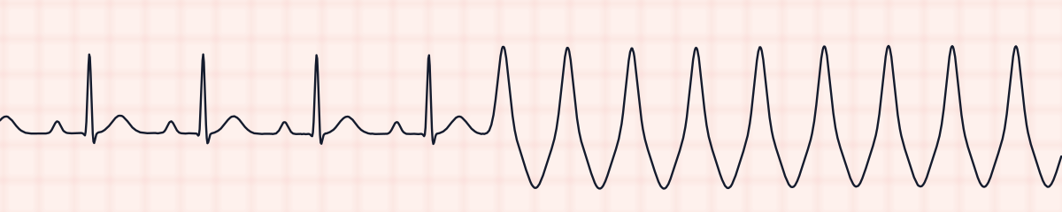
A rhythm is a set of rules, not a picture. These three strips all meet the rules for sinus rhythm converting to V-tach, with different numbers.
What sinus rhythm converting to V-tach gets mistaken for
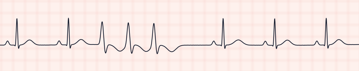
Sinus rhythm converting to V-tach vs. ventricular triplet (three PVCs in a row)
A triplet is three wide beats, after which the sinus rhythm returns and can be seen on the same strip. In a conversion the wide beats do not stop before the paper does.
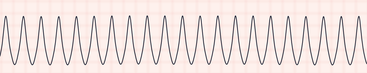
Sinus rhythm converting to V-tach vs. ventricular tachycardia
A strip that is ventricular tachycardia from edge to edge shows the same wide run without its beginning. When the sinus beats ahead of it are on the paper, the interpretation includes them, because they show what the patient’s rhythm was and the moment it changed.
Sinus rhythm converting to V-tach vs. torsades de pointes
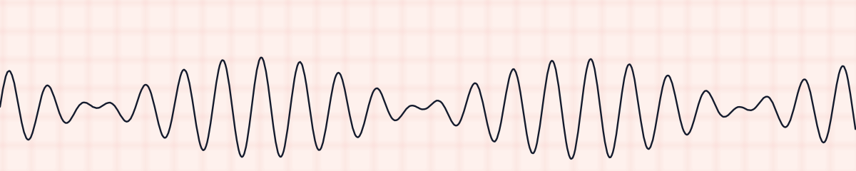
Torsades de pointes can also break out of a sinus rhythm. Its complexes swell and shrink and seem to twist about the baseline; the run on this kind of strip keeps one height and one shape from beat to beat.
Sinus rhythm converting to V-tach vs. supraventricular tachycardia (SVT)
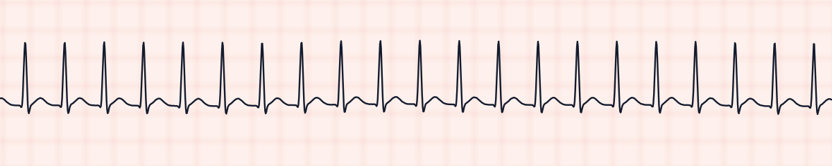
A sinus rhythm can jump to a fast rhythm with narrow complexes too. That is a supraventricular tachycardia. Measure the QRS of the fast beats: under 0.12 seconds points above the ventricles, 0.12 or more points to them.
Sinus rhythm converting to V-tach vs. ventricular couplet (paired PVCs)
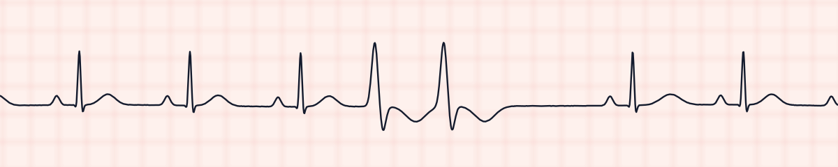
A couplet is over after two wide beats and a pause.
Practice: sinus rhythm converting to V-tach or a look-alike?
Five strips. Some show sinus rhythm converting to V-tach; the others are the rhythms most often confused with it.
Strip 1 of 5
Lead II · 25 mm/s · 6 s
Causes of sinus rhythm converting to V-tach
- A heart attack, or muscle that is short of blood supply
- An old heart attack that left scar in the ventricle
- Cardiomyopathy and heart failure with a low ejection fraction
- Low potassium or low magnesium
- A PVC that lands on the preceding T wave (R on T)
- Drugs that provoke ventricular arrhythmias, including digoxin in toxic amounts and some antiarrhythmics
- Low oxygen or severe acidosis
- Stimulants such as cocaine and amphetamines
Why it matters
This is the strip that sends a nurse to the bedside at once. At these rates the ventricles have little time to fill, so blood pressure can fall within seconds, and ventricular tachycardia can lose its pulse altogether or break down into ventricular fibrillation. Six seconds of paper cannot show how long the run will last: it may end by itself after a few beats or carry on. The monitor shows the rhythm; only the patient shows whether it is producing a pulse, and everything that follows depends on that.
What is done for sinus rhythm converting to V-tach
- Go to the patient immediately: are they responsive, are they breathing, is there a pulse?
- No pulse: call the code and start CPR. Without a pulse this rhythm is shockable, and the shock is given as soon as a defibrillator is at the bedside.
- A pulse is present: call for help, take a blood pressure, give oxygen if needed, make sure of IV access and bring the defibrillator to the bedside.
- Whether the patient is stable decides what the team does next: synchronized cardioversion when they are unstable, an antiarrhythmic drug when they are stable, as the provider orders.
- Print the strip that shows the onset and record the time. The first beat of the run helps explain what triggered it.
- Once the patient is safe, expect a 12-lead ECG and potassium and magnesium levels.
Study summary, not a protocol. Follow current American Heart Association guidelines and your facility’s policies. See the medical disclaimer.
Common mistakes with sinus rhythm converting to V-tach
- Watching the monitor to see whether it stops. The patient is checked first; the rhythm can be studied afterwards.
- Reporting ventricular tachycardia without mentioning the sinus rhythm before it or the time of onset.
- Calling a continuing run a “triplet” or “a few PVCs” because it began with one early beat.
- Mistaking movement artifact for ventricular tachycardia. With artifact, such as from toothbrushing or shivering, the patient’s narrow QRS complexes can still be found marching through the wide waves at the old rate.
- Assuming a patient who is awake cannot be in ventricular tachycardia. Some people keep a pulse and stay alert for a while.
Questions about sinus rhythm converting to V-tach
What does “sinus rhythm converting to ventricular tachycardia” mean?
That one strip shows both rhythms and the changeover between them: sinus beats first, then a run of fast, wide ventricular beats that continues. It is read as two rhythms in sequence, not as a single one.
How many wide beats in a row count as V-tach?
At least three, one after another, coming faster than 100 a minute. If the burst is over within 30 seconds without treatment it is labelled nonsustained. Lasting 30 seconds or more, or needing to be terminated because the patient is deteriorating, makes it sustained.
How can you tell where the V-tach starts?
Find the last QRS that is narrow and has a P wave ahead of it. The next complex is wide, comes sooner than a sinus beat would have, and has no P wave: that is the first beat of the tachycardia.
What should a nurse do first on seeing this strip?
Check the patient for responsiveness and a pulse. Pulseless ventricular tachycardia is treated as cardiac arrest with CPR and defibrillation; with a pulse, the team is called, vital signs are taken and treatment follows the patient’s condition.
Is a run of V-tach the same as sustained V-tach?
No. “Run” usually describes a short, self-ending burst seen in passing on the monitor. Sustained ventricular tachycardia keeps going. A six-second strip that ends in mid-run cannot tell the two apart, which is why the patient is assessed straight away.