AV blocks
2:1 AV Block on an EKG Strip
A 2:1 AV block is a second-degree heart block in which every other P wave fails to reach the ventricles. The strip shows two P waves for each QRS: one that is followed by a QRS, always after the same PR interval, and one that stands alone. The ventricles beat regularly, at exactly half the atrial rate.
Criteria from the American Heart Association guidelines and published ECG standards · Every strip checked by measurement · Sources and method · Updated November 15, 2026
- Rate
- Ventricular = half the atrial
- PR interval
- Constant on conducted beats
- QRS
- Narrow or wide
How to identify 2:1 AV block in five steps
Ask the same five questions of every strip. The right-hand column is what each one finds on the strip above.
1Rate
Count two rates. The ventricular rate is half the atrial rate: P waves at 80 a minute give QRS complexes at 40.
34 bpm; atrial 67
2Rhythm
Regular. The R-R intervals are even, and so are the P-P intervals.
Regular
3P waves
Two for each QRS, all of one shape and evenly spaced. The one that is not conducted often sits just behind the T wave, where it is easily taken for part of it.
2 per QRS
4PR interval
Identical on every conducted beat, and often normal. There is no second conducted beat in a row to compare it with.
0.19 s on conducted beats
5QRS
Narrow or wide. A wide QRS points toward a block below the AV node without settling the question.
0.07 s
What is happening in the heart
In any second-degree block some atrial impulses get through and some do not. In type I (Wenckebach) the AV node tires a little more with each beat, so the PR interval stretches until an impulse fails. In type II the fault lies lower, in the bundle of His or the bundle branches, and impulses fail with no warning. The two are told apart by comparing one conducted beat with the next. With 2:1 conduction that comparison is impossible, because each conducted beat is followed by a blocked one and never by another conducted beat. The strip proves that the block is second degree and how often it drops a beat. It cannot show which type it is.
More 2:1 AV block strips: no two are the same
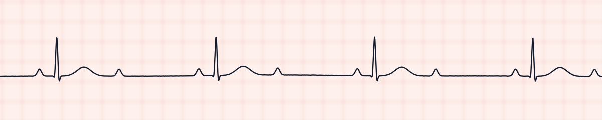
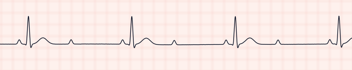
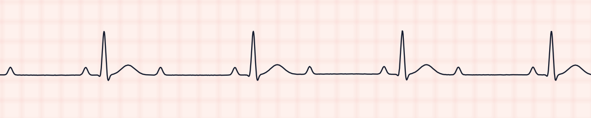
A rhythm is a set of rules, not a picture. These three strips all meet the rules for 2:1 AV block, with different numbers.
What 2:1 AV block gets mistaken for
2:1 AV block vs. second-degree AV block, type II (Mobitz II)
Mobitz II shows at least two conducted beats side by side with a matching PR interval before the one that is dropped. If a strip has such a pair anywhere on it, it is type II and not “2:1”.
2:1 AV block vs. second-degree AV block, type I (Wenckebach)
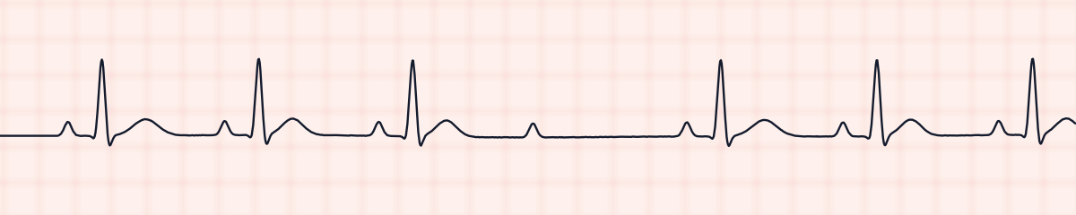
Wenckebach also has conducted beats next to each other, and across them the PR interval grows. A long recording of 2:1 block sometimes slips into 3:2 conduction for a cycle, and that one cycle gives the type away.
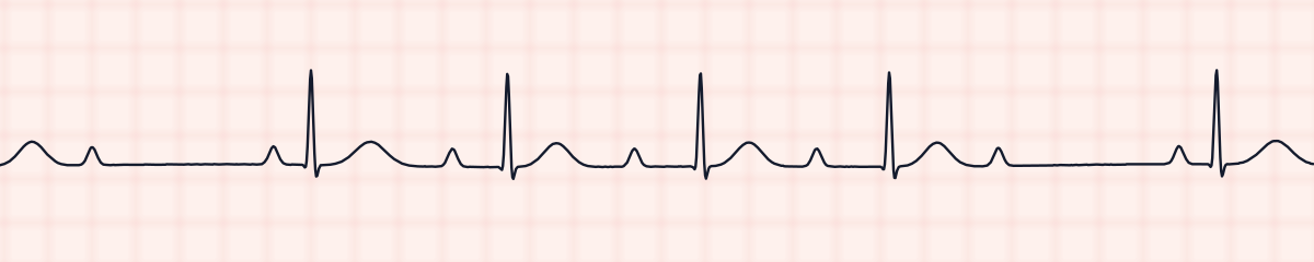
2:1 AV block vs. third-degree (complete) AV block
Complete heart block has more P waves than QRS complexes and a slow, even ventricular rate as well. Measure the PR interval on several beats. In 2:1 block it comes out the same each time; in complete block no two agree.
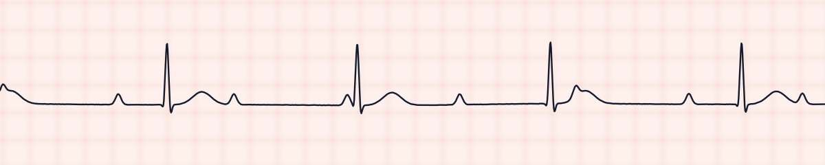
2:1 AV block vs. sinus bradycardia
When the second P wave hides in the T wave, the strip passes for sinus bradycardia at 35 to 45 a minute. Compare the T waves with one another and walk the P-P interval across the strip: a P wave lands on or just after every T wave.
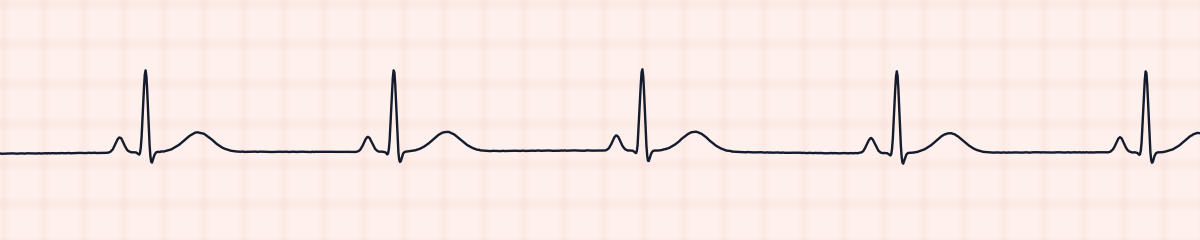
Practice: 2:1 AV block or a look-alike?
Five strips. Some show 2:1 AV block; the others are the rhythms most often confused with it.
Strip 1 of 5
Lead II · 25 mm/s · 6 s
Causes of 2:1 AV block
- Drugs that slow AV conduction: beta blockers, diltiazem, verapamil, digoxin
- An inferior heart attack, which usually affects the AV node itself
- An anterior heart attack, which can damage the conduction system below the node
- Scarring of the conduction system with age
- High vagal tone, during sleep or in trained athletes
- Recent heart surgery or a valve procedure
- Myocarditis and Lyme disease, less often
- A high potassium level
Why it matters
A 2:1 block halves the ventricular rate, and at 30 to 45 beats a minute many patients feel it: light-headedness, fatigue, breathlessness, a falling blood pressure. The second concern is where it may lead. A block of the type II kind can become complete heart block abruptly, and since the strip cannot say which kind this is, a new 2:1 block is handled with that possibility in mind until more is known. What helps the provider decide is the width of the QRS, a longer recording that catches the ratio changing, and the circumstances in which the block appeared.
What is done for 2:1 AV block
- Assess the patient: blood pressure, alertness, chest pain, shortness of breath.
- Run a long strip. A stretch of 3:2 conduction, if one turns up, reveals the type.
- Record the atrial rate, the ventricular rate, the PR interval and the QRS width, and name it second-degree AV block with 2:1 conduction.
- Look through the medication list for anything that slows the AV node, and check the most recent potassium.
- Report a new block to the provider, and keep pacing equipment close for a patient who has symptoms.
- A slow rate with symptoms is treated by the bradycardia pathway, as the provider directs.
Study summary, not a protocol. Follow current American Heart Association guidelines and your facility’s policies. See the medical disclaimer.
Common mistakes with 2:1 AV block
- Calling it Mobitz II because the PR interval is constant. It is constant in every 2:1 block, including those that are type I: there is only one conducted PR interval at a time to look at.
- Calling it sinus bradycardia after taking the extra P wave for a bump on the T wave.
- Calling it complete heart block because P waves outnumber QRS complexes two to one. Check whether the PR interval repeats.
- Writing down the ventricular rate alone. The atrial rate is the other half of the finding.
- Deciding the type from a single 6-second strip.
Questions about 2:1 AV block
Is 2:1 AV block Mobitz type 1 or type 2?
The strip alone cannot say. Either type can conduct 2:1. Naming the type takes two conducted beats in succession, to see whether the PR interval lengthens, and 2:1 conduction never provides them. The finding is reported as second-degree AV block with 2:1 conduction.
How do you recognize 2:1 AV block on an EKG?
By three things together: two evenly spaced P waves for each QRS, one unchanging PR interval on the beats that conduct, and a regular ventricular rhythm at half the atrial rate.
What helps to tell which type a 2:1 block is?
A longer recording in which the ratio changes, for instance to 3:2. Beyond that, a narrow QRS with a long PR interval favours type I, in the AV node, and a wide QRS with a normal PR interval favours type II, below it. These are pointers for the provider, not proof.
Is 2:1 AV block the same as high-grade AV block?
No. High-grade, or advanced, AV block means that two or more P waves in a row are not conducted, as in 3:1 conduction. In 2:1 block only every other P wave is lost.
Is 2:1 AV block dangerous?
It can be. The halved heart rate may produce symptoms, and a block of the type II kind can go on to complete heart block. A new 2:1 block is reported promptly and the patient is watched closely.