Supraventricular rhythms
Wolff-Parkinson-White (WPW) on an EKG Strip
Wolff-Parkinson-White (WPW) pattern is the EKG sign of an extra electrical connection between the atria and the ventricles. In sinus rhythm it shows three things together: a PR interval shorter than 0.12 seconds, a slurred start to the QRS called a delta wave, and a QRS that the slur widens to about 0.12 seconds or more.
Criteria from the American Heart Association guidelines and published ECG standards · Every strip checked by measurement · Sources and method · Updated November 15, 2026
- Rate
- 60–100
- PR interval
- < 0.12 s
- QRS
- ≥ 0.12 s, with a delta wave
How to identify WPW in five steps
Ask the same five questions of every strip. The right-hand column is what each one finds on the strip above.
1Rate
60 to 100 while the patient is in sinus rhythm.
91 bpm
2Rhythm
Regular while the patient stays in sinus rhythm.
Regular
3P waves
Upright, one per QRS, running almost straight into it.
1 per QRS
4PR interval
Shorter than 0.12 seconds.
0.11 s, short
5QRS
About 0.12 seconds or wider, with a slow slur at the foot of the upstroke.
0.14 s, with a delta wave
What is happening in the heart
Normally the AV node is the only electrical route from the atria to the ventricles, and it holds each impulse back for about a tenth of a second. In WPW a strand of muscle, the accessory pathway, bridges the two somewhere else and does not hold anything back. Part of a ventricle is therefore activated early, which shortens the PR interval, and it is activated slowly, cell to cell, which draws the sloping delta wave. A moment later the impulse that went through the AV node arrives and finishes the QRS with the usual sharp upstroke.
More WPW strips: no two are the same
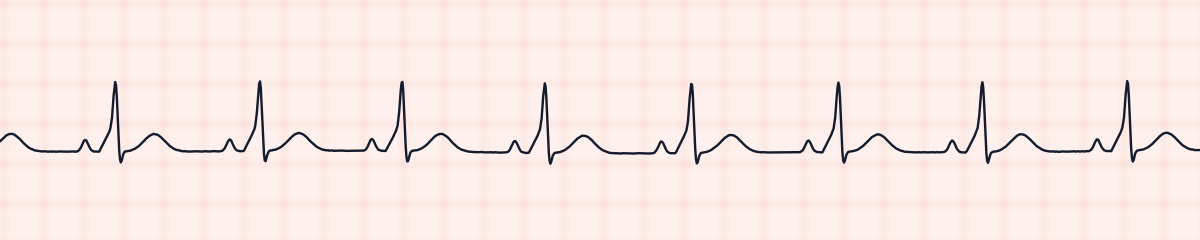
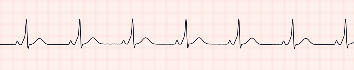
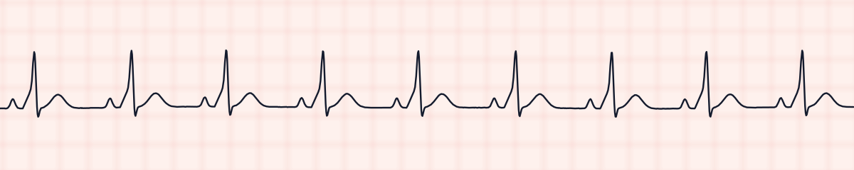
A rhythm is a set of rules, not a picture. These three strips all meet the rules for WPW, with different numbers.
What WPW gets mistaken for
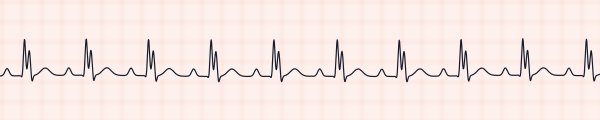
WPW vs. sinus rhythm with bundle branch block
A bundle branch block widens the end of the QRS and leaves the PR interval alone. Pre-excitation widens the beginning and shortens the PR. Put the calipers on the PR interval before anything else.
WPW vs. normal sinus rhythm
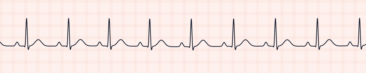
A quick look can pass WPW as normal, since it is regular and has P waves. The PR interval and the first 0.04 seconds of the QRS give it away.
WPW vs. accelerated junctional rhythm
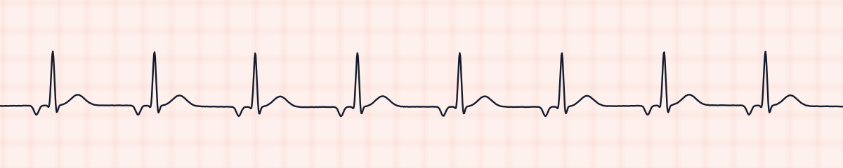
A junctional rhythm can also have a short PR interval, but its P wave is upside down and its QRS is narrow with a clean start.
Practice: WPW or a look-alike?
Five strips. Some show WPW; the others are the rhythms most often confused with it.
Strip 1 of 5
Lead II · 25 mm/s · 6 s
Causes of WPW
- An accessory pathway that has been present since birth
- It occasionally runs in families
- In a minority of cases it accompanies Ebstein anomaly or hypertrophic cardiomyopathy
- The pattern can come and go, because conduction over the pathway may be intermittent
Why it matters
The pattern by itself produces no symptoms, and many people learn of it from a routine EKG. The risk lies in the tachycardias the pathway makes possible. An impulse can circle down the AV node and back up the pathway, causing sudden episodes of SVT. More dangerous is atrial fibrillation: the pathway has no built-in delay, so it can pass very rapid, irregular impulses to the ventricles, and a fast, irregular, wide-complex rhythm in a patient with WPW is an emergency. Drugs that slow only the AV node are avoided in that situation because they can speed conduction over the pathway.
What is done for WPW
- Document the pattern and report it when it has not been noted before.
- Ask about palpitations, a racing heart, light-headedness or fainting.
- Expect a 12-lead EKG and a referral to cardiology or electrophysiology.
- Catheter ablation of the pathway is the usual definitive treatment for patients who have symptoms.
- For a tachycardia in a patient known to have WPW, the choice of drug belongs to the provider; an unstable patient is cardioverted.
Study summary, not a protocol. Follow current American Heart Association guidelines and your facility’s policies. See the medical disclaimer.
Common mistakes with WPW
- Skipping the PR measurement because P waves are present and the rhythm is regular.
- Mistaking the delta wave for the tail of the P wave or for a Q wave.
- Calling the wide QRS a bundle branch block without checking where the extra width lies.
- Using “WPW syndrome” for the EKG pattern alone. The syndrome means the pattern plus episodes of tachycardia.
Questions about WPW
What is a delta wave?
A delta wave is the slurred, slanting start of the QRS in pre-excitation. Instead of leaving the baseline sharply, the upstroke climbs slowly for the first few hundredths of a second and then steepens.
What are the three EKG signs of WPW?
A short PR interval (under 0.12 seconds), a delta wave, and a QRS that is wider than normal because of the delta wave.
Is WPW dangerous?
Most people with the pattern never have a serious event. The concern is a fast arrhythmia, above all atrial fibrillation conducted over the accessory pathway, which can deteriorate into ventricular fibrillation.
What is the difference between WPW pattern and WPW syndrome?
The pattern is the EKG finding only. The syndrome is diagnosed when a person with that finding also has symptomatic tachycardia.