Ventricular rhythms
Ventricular tachycardia (V-tach) EKG strip
Ventricular tachycardia (VT or V-tach) is a fast rhythm that starts in the ventricles. On an EKG strip it is a run of wide QRS complexes (0.12 seconds or more) at more than 100 beats per minute, usually 150 to 250, regular, with no P waves in front of them. Three or more such beats in a row count as VT.
Criteria from the American Heart Association guidelines and published ECG standards · Every strip checked by measurement · Sources and method · Updated November 15, 2026
- Rate
- 100–250
- PR interval
- None
- QRS
- ≥ 0.12 s
How to identify ventricular tachycardia in five steps
Ask the same five questions of every strip. The right-hand column is what each one finds on the strip above.
1Rate
More than 100 per minute, commonly 150 to 250.
208 bpm
2Rhythm
Regular, or very nearly so.
Regular
3P waves
Not visible in front of the QRS complexes.
None
4PR interval
None.
None
5QRS
0.12 s or wider, all alike in monomorphic VT, with the T wave pointing the opposite way.
0.16 s, wide
What is happening in the heart
An irritable area or a short circuit in the ventricular muscle, often at the edge of scar from an old heart attack, takes over as pacemaker and fires rapidly. The impulse spreads through the muscle cell to cell, which is slow and produces a wide, bizarre QRS. The atria may still be beating on their own, but their P waves are lost among the large ventricular complexes.
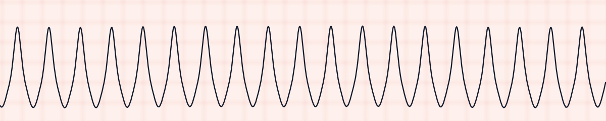
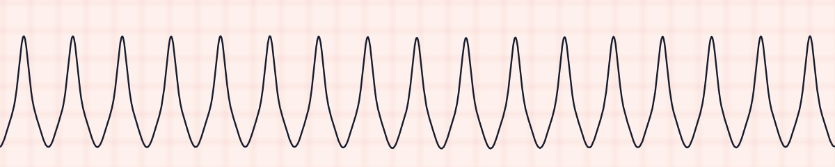
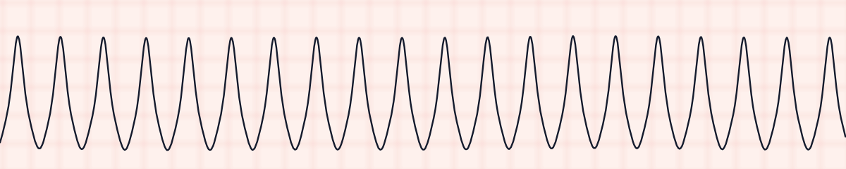
More ventricular tachycardia strips: no two are the same
A rhythm is a set of rules, not a picture. These three strips all meet the rules for ventricular tachycardia, with different numbers.
What ventricular tachycardia gets mistaken for
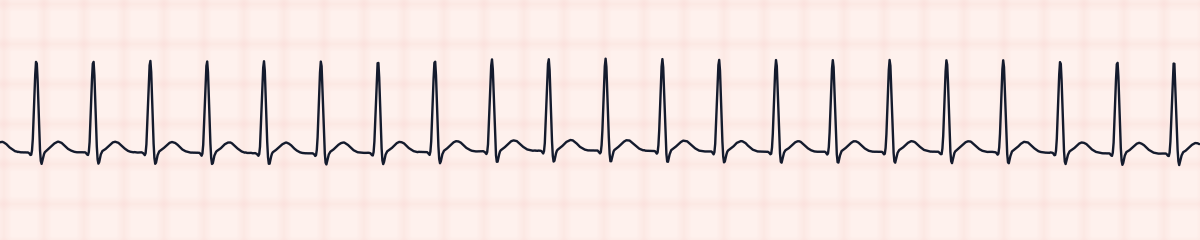
Ventricular tachycardia vs. supraventricular tachycardia (SVT)
Both are fast and regular. SVT has a narrow QRS. A wide regular tachycardia is treated as ventricular tachycardia unless someone proves otherwise.
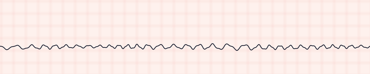
Ventricular tachycardia vs. ventricular fibrillation
Ventricular tachycardia has organized complexes that repeat with the same shape and spacing. Ventricular fibrillation has no repeating shape at all.
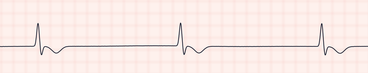
Ventricular tachycardia vs. idioventricular rhythm
The same wide complexes at a slow rate. Under 40 is an idioventricular rhythm, 40 to 100 is accelerated idioventricular rhythm, and over 100 is ventricular tachycardia.
Practice: ventricular tachycardia or a look-alike?
Five strips. Some show ventricular tachycardia; the others are the rhythms most often confused with it.
Strip 1 of 5
Lead II · 25 mm/s · 6 s
Causes of ventricular tachycardia
- Myocardial infarction and ischemia
- Scar from a previous heart attack
- Cardiomyopathy and heart failure
- Low potassium or low magnesium
- Drugs that prolong the QT interval
- Stimulants
Why it matters
Ventricular tachycardia can look exactly the same on the monitor in a patient who is talking to you and in one who is in cardiac arrest. The fast rate leaves little time for the ventricles to fill, so blood pressure can collapse, and the rhythm can break down into ventricular fibrillation. The first action is always the same: check for a pulse.
What is done for ventricular tachycardia
- Check for a pulse and responsiveness first.
- No pulse: this is cardiac arrest. Start CPR and defibrillate, exactly as for ventricular fibrillation.
- Pulse but unstable (low blood pressure, altered mental status, chest pain, shock): synchronized cardioversion.
- Pulse and stable: an antiarrhythmic such as amiodarone, with expert consultation.
- Check potassium and magnesium, and look for the cause.
Study summary, not a protocol. Follow current American Heart Association guidelines and your facility’s policies. See the medical disclaimer.
Common mistakes with ventricular tachycardia
- Treating the monitor instead of the patient. The pulse check decides everything.
- Calling it SVT without measuring the QRS.
- Reacting to artifact. Tooth brushing or shivering can imitate VT; a patient who is awake and comfortable with a normal pulse is not in sustained VT.
Questions about ventricular tachycardia
What does V-tach look like on an EKG strip?
A fast, regular run of wide QRS complexes with no P waves in front of them. The complexes are tall, broad and look alike, and the rate is usually 150 to 250 per minute.
Is ventricular tachycardia shockable?
Pulseless ventricular tachycardia is a shockable rhythm and is defibrillated. Ventricular tachycardia with a pulse in an unstable patient is treated with synchronized cardioversion instead.
How many beats make ventricular tachycardia?
Three or more ventricular beats in a row at a rate above 100 per minute. A run lasting more than 30 seconds, or one that causes collapse, is called sustained.
What is the difference between monomorphic and polymorphic VT?
In monomorphic VT every QRS has the same shape. In polymorphic VT the shape and height keep changing. Torsades de pointes is a polymorphic VT linked to a long QT interval and is treated with magnesium.