Paced rhythms
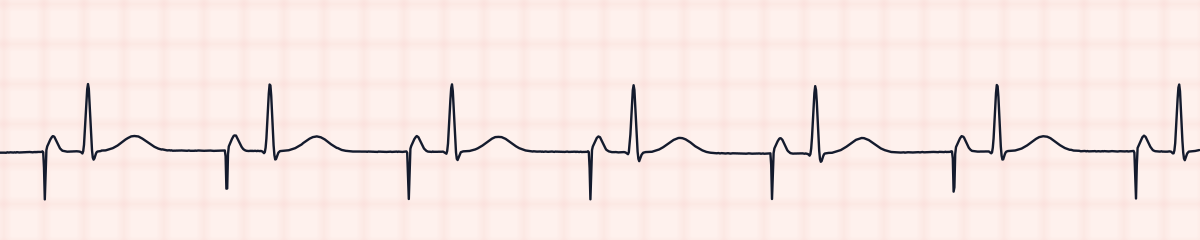
Atrial paced rhythm EKG strip
An atrial paced rhythm is produced by a pacemaker stimulating the atria. On an EKG strip each beat starts with a pacing spike, followed at once by a P wave, then a normal PR interval and a narrow QRS. The rhythm is regular at the pacemaker’s set rate.
Criteria from the American Heart Association guidelines and published ECG standards · Every strip checked by measurement · Sources and method · Updated November 15, 2026
- Rate
- Set rate, often 60–80
- PR interval
- Normal, spike to QRS
- QRS
- < 0.12 s
How to identify an atrial paced rhythm in five steps
Ask the same five questions of every strip. The right-hand column is what each one finds on the strip above.
1Rate
The set rate, commonly 60 to 80, and steady.
67 bpm
2Rhythm
Regular.
Regular
3P waves
One per beat, each immediately after a spike. Its shape may differ a little from a sinus P wave.
Each after a pacing spike
4PR interval
Measured from the spike to the QRS; normal and constant.
0.20 s
5QRS
Under 0.12 s.
0.08 s
What is happening in the heart
The pacing lead sits in the right atrium and does the job of the SA node. Its pulse triggers the atria, which draws the P wave. From there the impulse passes through the patient’s own AV node and bundle branches, so the ventricles are activated normally and the QRS is narrow. Atrial pacing therefore only works when AV conduction is intact.
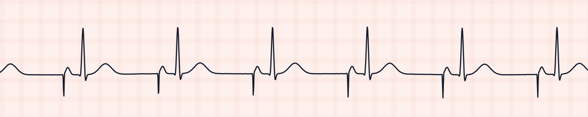
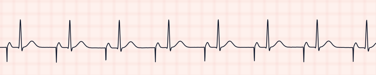
More atrial paced rhythm strips: no two are the same
A rhythm is a set of rules, not a picture. These three strips all meet the rules for an atrial paced rhythm, with different numbers.
What an atrial paced rhythm gets mistaken for
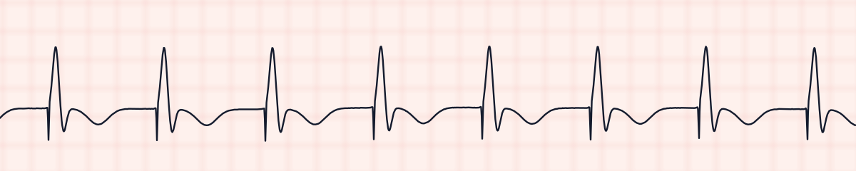
Atrial paced rhythm vs. ventricular paced rhythm
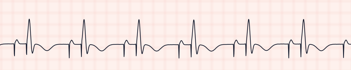
In ventricular pacing the spike is attached to the QRS, which is wide. In atrial pacing the spike is attached to the P wave, and the QRS that follows is narrow.
Atrial paced rhythm vs. AV sequential paced rhythm
AV pacing has two spikes per beat and a wide QRS. Atrial pacing has one spike per beat and a narrow QRS.
Atrial paced rhythm vs. normal sinus rhythm
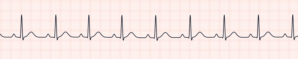
Take the spikes away and the two look the same. If the spike is small it can be missed, so look at the very start of each P wave.
Practice: an atrial paced rhythm or a look-alike?
Five strips. Some show an atrial paced rhythm; the others are the rhythms most often confused with it.
Strip 1 of 5
Lead II · 25 mm/s · 6 s
Why a patient is paced this way
- Sinus node dysfunction (sick sinus syndrome)
- Symptomatic sinus bradycardia
- Temporary pacing wires after cardiac surgery
- Sinus pauses
Why it matters
Atrial pacing keeps the normal sequence of the heartbeat: atria first, then ventricles through the natural pathway. That preserves the atrial contribution to filling and a normal narrow QRS. The checks are that every spike produces a P wave, and that every P wave is still being conducted to the ventricles.
What to check with an atrial paced rhythm
- Confirm atrial capture: a P wave after every atrial spike.
- Confirm that each P wave is followed by a QRS.
- Check a pulse against the monitor.
- Know the set rate and report a rate below it.
- Report spikes with no P wave after them, and P waves that stop conducting.
Study summary, not a protocol. Follow current American Heart Association guidelines and your facility’s policies. See the medical disclaimer.
Common mistakes with an atrial paced rhythm
- Missing the spike and reporting normal sinus rhythm.
- Calling it a ventricular paced rhythm without noticing the QRS is narrow.
- Not noticing a new AV block developing behind the pacing.
Questions about an atrial paced rhythm
What does an atrial paced rhythm look like?
A pacing spike, then a P wave, then a normal narrow QRS. The spike comes before the P wave, not before the QRS.
Why is the QRS narrow in atrial pacing?
Only the atria are paced. The impulse reaches the ventricles through the normal AV node and bundle branches, so they are activated quickly and together, as in a sinus beat.
What is the difference between atrial and ventricular pacing?
Atrial pacing puts the spike before the P wave and gives a narrow QRS. Ventricular pacing puts the spike before the QRS, which is wide because the impulse starts in the ventricle.