Arrest rhythms
Pulseless electrical activity (PEA) rhythm strip
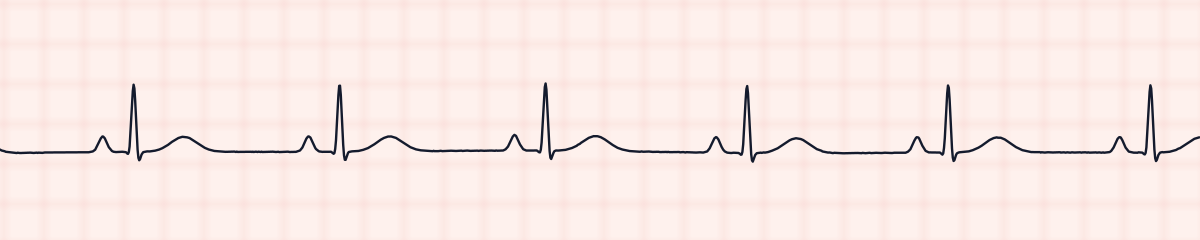
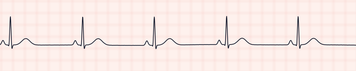
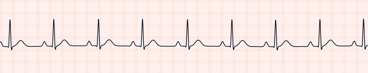
Pulseless electrical activity (PEA) is a cardiac arrest in which the monitor shows an organized rhythm but the patient has no pulse. It is not one pattern on the EKG strip: any organized rhythm, including one that looks like normal sinus rhythm, is PEA if the heart is not producing a pulse. It is not shockable.
Criteria from the American Heart Association guidelines and published ECG standards · Every strip checked by measurement · Sources and method · Updated November 15, 2026
- Rate
- Any
- PR interval
- Any
- QRS
- Any
How to identify PEA in five steps
Ask the same five questions of every strip. The right-hand column is what each one finds on the strip above.
1Rate
Any rate. It is often slow, and it tends to slow further as the arrest goes on.
81 bpm
2Rhythm
Any organized rhythm, regular or not.
Regular
3P waves
May be present or absent, depending on the rhythm.
1 per QRS
4PR interval
Whatever the underlying rhythm has.
0.13 s
5QRS
Narrow or wide. A wide, slow PEA usually means a more damaged heart.
0.09 s
What is happening in the heart
The electrical system is still working, so impulses form and travel and the monitor draws complexes. But the heart muscle does not turn those signals into an effective contraction, or it contracts with no blood to pump. Something outside the electrical system is preventing output: too little blood volume, too little oxygen, pressure squeezing the heart, or a clot blocking flow.
Three strips that could all be PEA
Each of these is an organized rhythm. With a pulse it is sinus rhythm. Without one it is PEA. The strip cannot tell you which.
What PEA gets mistaken for
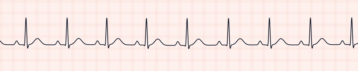
PEA vs. normal sinus rhythm
There may be no difference at all on the strip. The difference is at the bedside: a patient in normal sinus rhythm has a pulse. This is why you never name a rhythm as “fine” without looking at the patient.
PEA vs. asystole
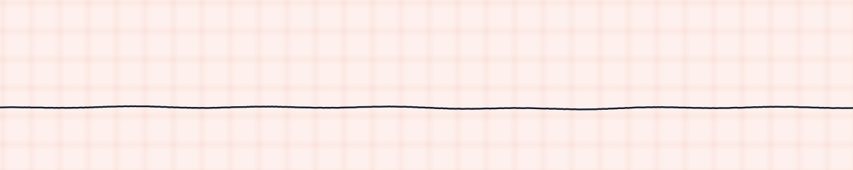
Both are non-shockable arrest rhythms and share one treatment pathway. Asystole is a flat line; PEA has organized complexes.
Causes of PEA
- Hypovolemia (severe blood or fluid loss), the most common reversible cause
- Hypoxia
- Hydrogen ion excess (acidosis)
- Hypokalemia or hyperkalemia
- Hypothermia
- Tension pneumothorax
- Tamponade (cardiac)
- Toxins and overdose
- Thrombosis: pulmonary embolism or heart attack
Why it matters
PEA is the reason for the rule “treat the patient, not the monitor.” A rhythm that looks reassuring can belong to someone in cardiac arrest. Survival depends almost entirely on finding and reversing the cause, which is why the list of Hs and Ts is memorized.
What is done for PEA
- Confirm there is no pulse, and call for help.
- Start high-quality CPR immediately.
- Give epinephrine during the resuscitation.
- Do not defibrillate. PEA is not a shockable rhythm.
- Work through the Hs and Ts while CPR continues, and treat any cause you find.
- Recheck the rhythm and pulse at the scheduled pauses; PEA can change to a shockable rhythm.
Study summary, not a protocol. Follow current American Heart Association guidelines and your facility’s policies. See the medical disclaimer.
Common mistakes with PEA
- Looking at an organized rhythm on the monitor and not checking for a pulse.
- Shocking PEA because the patient is in arrest. Only VF and pulseless VT are shocked.
- Running the resuscitation without looking for a cause. CPR and epinephrine buy time; reversing the cause is the treatment.
Questions about PEA
Can you identify PEA from an EKG strip?
No. PEA is defined by two things together: an organized rhythm on the monitor and no pulse in the patient. The strip shows only the first. This is why PEA is not asked as an identify-the-strip question on this site.
What rhythm is PEA?
Any organized rhythm without a pulse: sinus rhythm, a slow junctional rhythm, an idioventricular rhythm and others. Ventricular fibrillation, pulseless ventricular tachycardia and asystole are named separately and are not called PEA.
Is PEA shockable?
No. PEA and asystole are the non-shockable arrest rhythms. They are treated with CPR, epinephrine and correction of the underlying cause.
What are the Hs and Ts?
A memory aid for the reversible causes of cardiac arrest: hypovolemia, hypoxia, hydrogen ion (acidosis), hypo- or hyperkalemia, hypothermia; tension pneumothorax, tamponade, toxins, and thrombosis in the lungs or the heart.