Supraventricular rhythms
Multifocal Atrial Tachycardia (MAT) on an EKG Strip
Multifocal atrial tachycardia (MAT) is a fast, irregular rhythm driven by several competing sites in the atria. It is recognized by a rate above 100 beats a minute, P waves of three or more shapes, and PR and R-R intervals that keep changing. It is seen mostly in people who are acutely ill with lung disease.
Criteria from the American Heart Association guidelines and published ECG standards · Every strip checked by measurement · Sources and method · Updated November 15, 2026
- Rate
- > 100
- PR interval
- Changes with the P wave
- QRS
- < 0.12 s
How to identify MAT in five steps
Ask the same five questions of every strip. The right-hand column is what each one finds on the strip above.
1Rate
Over 100, usually 100 to 150.
~110 bpm
2Rhythm
Irregularly irregular.
Irregular
3P waves
Three or more shapes, one ahead of each QRS, with flat baseline in between.
Three shapes, 1 per QRS
4PR interval
Different from one beat to the next.
Changes with the P wave
5QRS
Narrow, unless the patient also has a bundle branch block.
0.08 s
What is happening in the heart
In MAT a number of irritable spots in the atrial muscle fire on their own, each at its own pace, and whichever fires first sets off the next heartbeat. Since every spot lies somewhere different, every spot produces its own P wave shape and its own PR interval, and because nothing coordinates them the beats arrive at uneven intervals. Low oxygen, stretched atria and stimulant drugs all make atrial cells more likely to behave this way.
More MAT strips: no two are the same
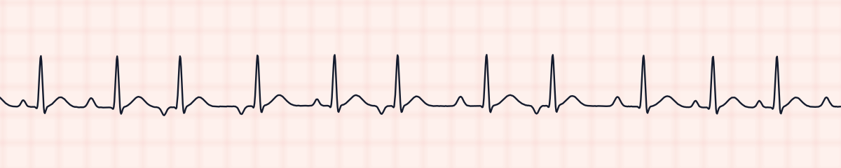
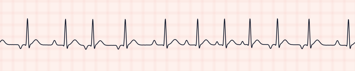
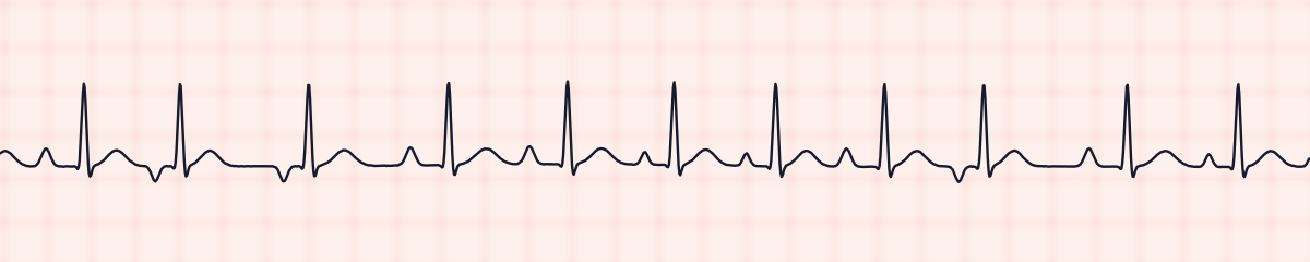
A rhythm is a set of rules, not a picture. These three strips all meet the rules for MAT, with different numbers.
What MAT gets mistaken for
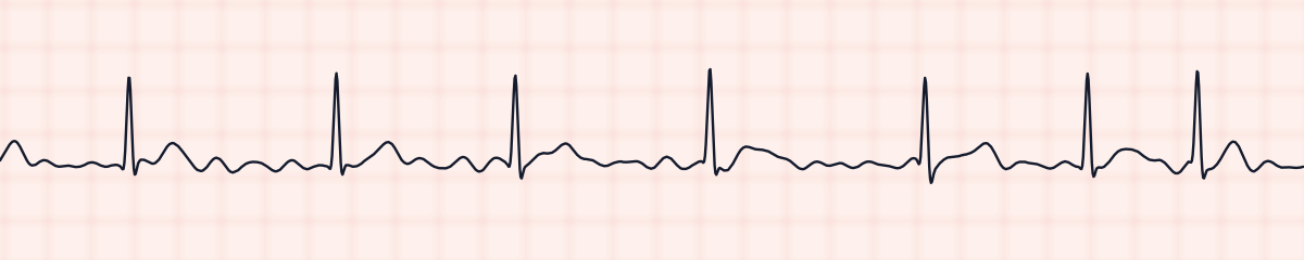
MAT vs. atrial fibrillation
Both are fast and irregularly irregular, and MAT is very often misread as atrial fibrillation. Fibrillation has a wavy baseline and no true P waves. MAT has a distinct P wave ahead of each QRS and a flat line between beats.
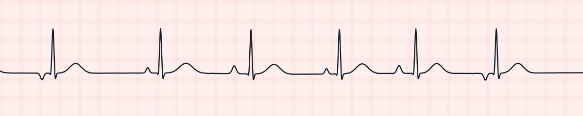
MAT vs. wandering atrial pacemaker
A wandering atrial pacemaker shows the same changing P waves at fewer than 100 beats a minute.
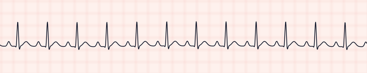
MAT vs. sinus tachycardia
Sinus tachycardia is fast and has P waves, but it is regular and its P waves are identical.
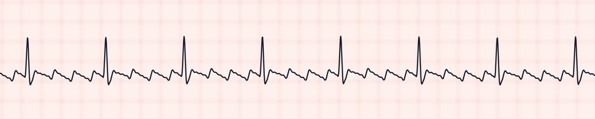
MAT vs. atrial flutter
Flutter with a varying block is irregular too, but its atrial waves are a uniform sawtooth at about 300 a minute.
Practice: MAT or a look-alike?
Five strips. Some show MAT; the others are the rhythms most often confused with it.
Strip 1 of 5
Lead II · 25 mm/s · 6 s
Causes of MAT
- A flare of COPD or other severe lung disease, by far the most common setting
- Low blood oxygen
- Pneumonia or sepsis
- Heart failure
- Low potassium or low magnesium
- Theophylline and other stimulant bronchodilators
- Pulmonary embolism
Why it matters
MAT is a marker of how sick the patient is more than a problem in itself. The fast rate adds to the work of a heart that is already short of oxygen, but the rhythm usually settles only when the illness behind it is treated. Mistaking it for atrial fibrillation leads to the wrong plan: electrical cardioversion does not work for MAT, and anticoagulation is not indicated for it.
What is done for MAT
- Assess breathing and oxygen saturation before anything else; the lungs are usually the source.
- Give oxygen as prescribed while the underlying illness is treated.
- Expect potassium and magnesium to be measured and corrected.
- Rate-slowing medication is sometimes ordered when the rate itself is causing trouble.
- Cardioversion is not used: there is no single circuit for a shock to interrupt.
Study summary, not a protocol. Follow current American Heart Association guidelines and your facility’s policies. See the medical disclaimer.
Common mistakes with MAT
- Reporting atrial fibrillation without checking the baseline for P waves.
- Not comparing the P waves with one another. They are easy to see and easy to assume alike.
- Measuring one PR interval and writing it down as if it applied to the whole strip.
- Treating the heart rate as the main problem when the cause is respiratory.
Questions about MAT
What are the EKG criteria for multifocal atrial tachycardia?
A rate above 100 beats a minute, at least three different P wave shapes in one lead, and varying P-P, PR and R-R intervals, with a flat baseline between the P waves.
How do you tell MAT from atrial fibrillation?
Look between the QRS complexes. In MAT a clear P wave comes before each one, although the shapes differ. In atrial fibrillation there are none, only a fine, wavy baseline.
What causes MAT?
It is strongly linked to serious lung disease, especially COPD exacerbations, and to low oxygen levels. Electrolyte problems, heart failure and theophylline also contribute.
How is MAT treated?
By treating what is behind it: improving oxygenation, treating infection, correcting magnesium and potassium. Drugs to slow the rate come second, and cardioversion is ineffective.