Ventricular rhythms
Premature ventricular contractions (PVCs) on an EKG strip
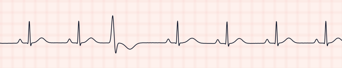
A premature ventricular contraction (PVC) is a single beat that starts early in the ventricles. On an EKG strip it is easy to spot: it comes before the next normal beat is due, has no P wave in front of it, is wide (0.12 seconds or more) and oddly shaped, and has a T wave pointing in the opposite direction. A full compensatory pause follows.
Criteria from the American Heart Association guidelines and published ECG standards · Every strip checked by measurement · Sources and method · Updated November 15, 2026
- Rate
- Underlying rhythm
- PR interval
- None on the early beat
- QRS
- ≥ 0.12 s on the early beat
How to identify PVCs in five steps
Ask the same five questions of every strip. The right-hand column is what each one finds on the strip above.
1Rate
That of the underlying rhythm. Count every QRS, including the PVCs, for the rate on the monitor.
~70 bpm
2Rhythm
An even sinus rhythm broken by a wide early beat and the long pause that follows it.
Regular, with early beats
3P waves
Normal on the sinus beats. None before the PVC.
None on the early beat
4PR interval
Normal on the sinus beats. Not measurable on the PVC.
0.13 s on sinus beats
5QRS
Narrow on the sinus beats. 0.12 s or wider and bizarre on the PVC.
0.08 s; early beat 0.15 s
What is happening in the heart
An irritable spot in the ventricular muscle fires on its own. The impulse spreads from cell to cell across the ventricles instead of down the fast conduction pathway, which takes longer and produces a wide, distorted QRS. The SA node is not disturbed and keeps its own time, but its next impulse arrives while the ventricles are still recovering and is wasted. The beat after that comes exactly on schedule, so the gap spanning the PVC equals two normal cycles.
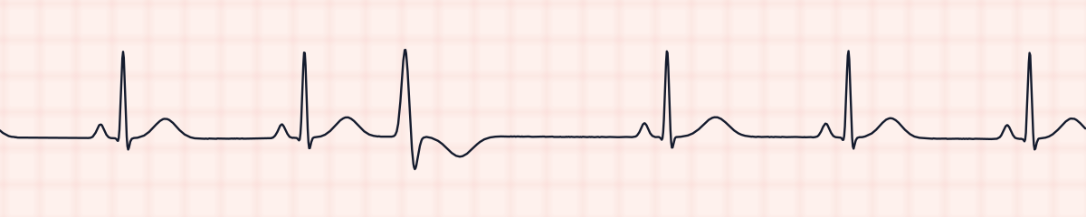
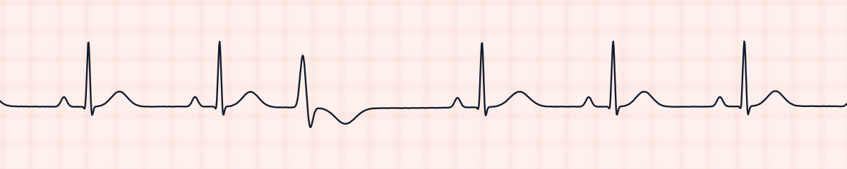
More PVC strips: no two are the same
A rhythm is a set of rules, not a picture. These three strips all meet the rules for PVCs, with different numbers.
What PVCs get mistaken for
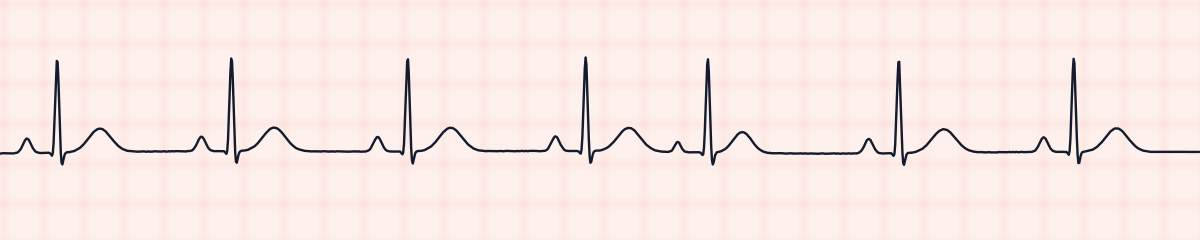
PVCs vs. premature atrial contractions (PACs)
A PAC is early too, but it has a P wave and a narrow QRS that looks like the patient’s other beats, and the pause after it is shorter. A PVC is wide and has no P wave.
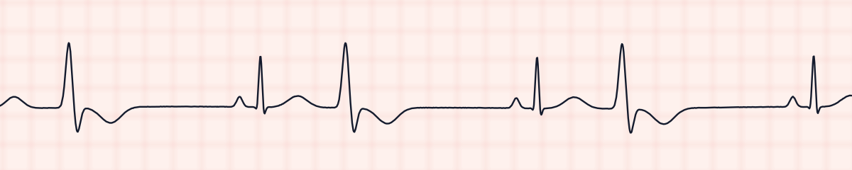
PVCs vs. ventricular bigeminy
Isolated PVCs come now and then with several normal beats between them. If every second beat is a PVC, the rhythm is ventricular bigeminy.
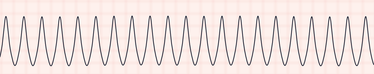
PVCs vs. ventricular tachycardia
One PVC is one wide beat. Three or more in a row at a rate over 100 is ventricular tachycardia.
Practice: PVCs or a look-alike?
Five strips. Some show PVCs; the others are the rhythms most often confused with it.
Strip 1 of 5
Lead II · 25 mm/s · 6 s
Causes of PVCs
- Hypoxia
- Myocardial ischemia and infarction
- Low potassium or low magnesium
- Caffeine, nicotine, alcohol and other stimulants
- Stress, anxiety and lack of sleep
- Heart failure and cardiomyopathy
- Digoxin toxicity
- Sometimes no cause at all, in a healthy heart
Why it matters
Occasional PVCs occur in healthy people and are usually harmless. They matter when they signal an irritable ventricle that could go on to ventricular tachycardia or fibrillation. The warning signs are PVCs that are frequent or increasing, that come in pairs or runs, that have more than one shape, that fall on the T wave of the beat before, or that appear during a heart attack.
What is done for PVCs
- Assess the patient, and check a pulse: a PVC may not produce one, so the pulse can be slower than the monitor rate.
- Check oxygen saturation, and potassium and magnesium results.
- Count how many occur per minute and note whether they are alike or of different shapes.
- Report new, frequent, paired or multiform PVCs, and any that land on a T wave.
- Treatment is aimed at the cause. Antiarrhythmic drugs are not given routinely for PVCs alone.
Study summary, not a protocol. Follow current American Heart Association guidelines and your facility’s policies. See the medical disclaimer.
Common mistakes with PVCs
- Ignoring the pattern. One PVC is rarely a problem; the trend over minutes and hours is what matters.
- Calling a wide early beat a PAC without checking for a P wave.
- Taking the heart rate from the monitor alone when many beats are PVCs that may not perfuse.
Questions about PVCs
What does a PVC look like on an EKG strip?
An early beat that is wide and bizarre compared with the normal beats, with no P wave before it and a T wave pointing the opposite way from the QRS. It is followed by a pause before the normal rhythm continues.
What is a compensatory pause?
The pause after a PVC. The SA node is not reset, so the next normal beat arrives exactly when it would have anyway. Measured from the beat before the PVC to the beat after it, the interval is exactly two normal R-R intervals.
When are PVCs dangerous?
When they are frequent (often quoted as more than six a minute), come in pairs or runs, have different shapes, fall on the preceding T wave, or occur with a heart attack or symptoms. These suggest a higher risk of ventricular tachycardia.
What is the difference between unifocal and multifocal PVCs?
Unifocal PVCs all look the same because they come from one spot. Multifocal PVCs have different shapes because they come from several spots, which indicates a more irritable ventricle.