Supraventricular rhythms
Atrial Fibrillation With PVCs on an EKG Strip
Atrial fibrillation with PVCs is a strip with two findings at once. The underlying rhythm is atrial fibrillation: no P waves, a wavy baseline and narrow QRS complexes at irregularly irregular intervals. Scattered through it are premature ventricular contractions, single beats that are wide, shaped differently from the rest and followed by a pause.
Criteria from the American Heart Association guidelines and published ECG standards · Every strip checked by measurement · Sources and method · Updated November 15, 2026
- Rate
- Count every QRS
- PR interval
- None
- QRS
- Narrow, with wide PVCs
How to identify atrial fibrillation with PVCs in five steps
Ask the same five questions of every strip. The right-hand column is what each one finds on the strip above.
1Rate
Count every QRS in six seconds, the PVCs included, and multiply by ten.
~100 bpm
2Rhythm
Irregularly irregular from one end of the strip to the other.
Irregularly irregular
3P waves
Absent, replaced by a fine, wavy baseline.
None
4PR interval
Cannot be measured.
Not measurable
5QRS
Narrow on the conducted beats; 0.12 seconds or wider on the PVCs.
0.08 s; PVC 0.16 s
What is happening in the heart
The two findings have separate sources. The atria are fibrillating and send a disorganized stream of impulses toward the AV node, which lets them through at unpredictable intervals; those beats use the normal pathway and are narrow. Independently, an irritable spot in a ventricle fires now and then. Its impulse spreads slowly from one muscle cell to the next, drawing a wide QRS with a T wave that points the opposite way, and it leaves the ventricles briefly unable to respond, so a pause follows.
More atrial fibrillation with PVCs strips: no two are the same
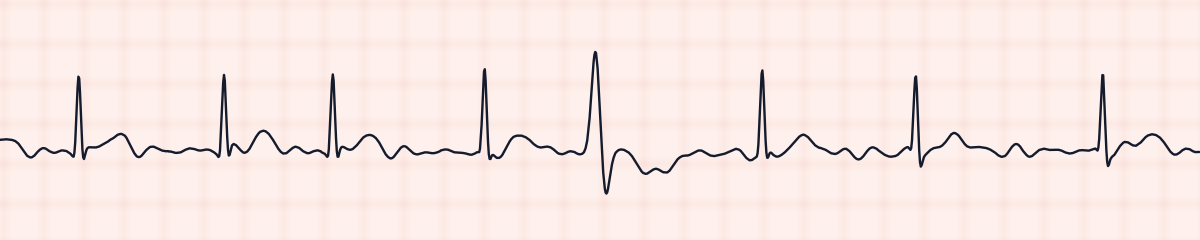
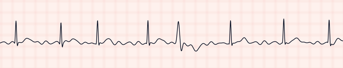
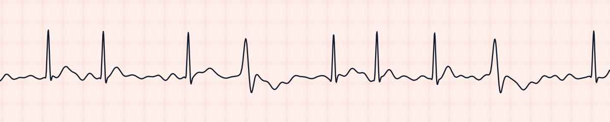
A rhythm is a set of rules, not a picture. These three strips all meet the rules for atrial fibrillation with PVCs, with different numbers.
What atrial fibrillation with PVCs get mistaken for
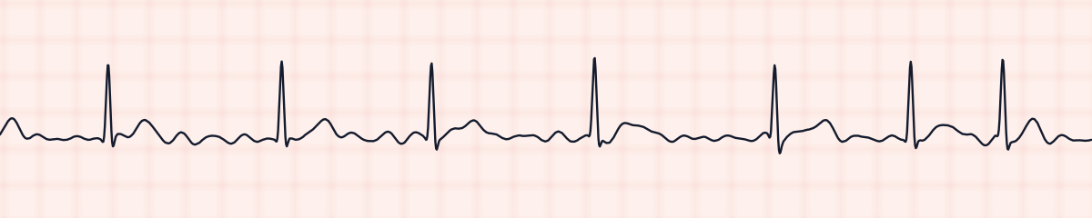
Atrial fibrillation with PVCs vs. atrial fibrillation
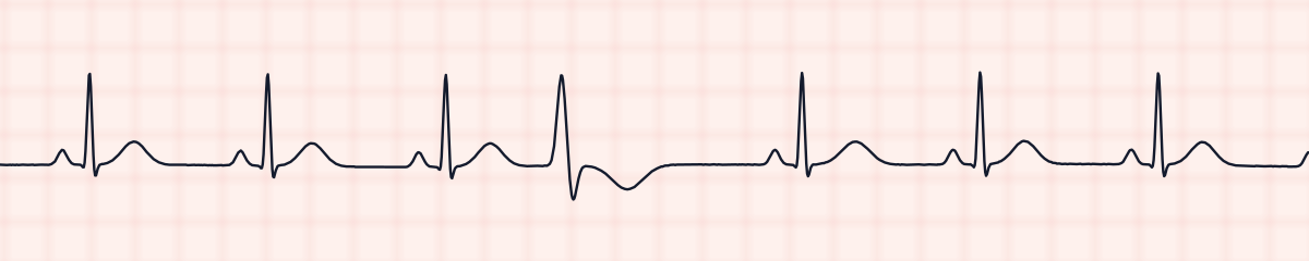
In plain atrial fibrillation the QRS complexes all look alike. One beat that is clearly wider and differently shaped makes it atrial fibrillation with PVCs.
Atrial fibrillation with PVCs vs. premature ventricular contractions (PVCs)
PVCs in sinus rhythm interrupt a regular rhythm that has P waves. Here the background is irregular and has none.
Atrial fibrillation with PVCs vs. ventricular tachycardia
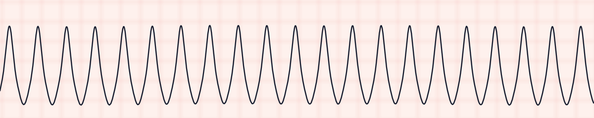
Three or more wide beats in a row are no longer isolated PVCs but a run of ventricular tachycardia.
Practice: atrial fibrillation with PVCs or a look-alike?
Five strips. Some show atrial fibrillation with PVCs; the others are the rhythms most often confused with it.
Strip 1 of 5
Lead II · 25 mm/s · 6 s
Causes of atrial fibrillation with PVCs
- Heart failure or cardiomyopathy, which predispose to both findings
- Coronary artery disease
- Low potassium or low magnesium, often from diuretics
- Digoxin toxicity: new PVCs in a patient who takes digoxin for atrial fibrillation are a warning sign
- Low oxygen
- Stimulants, including caffeine and some inhalers
- An overactive thyroid
Why it matters
Occasional PVCs add little risk to atrial fibrillation. They deserve attention when they are new, frequent or arrive in pairs and runs, because that points to an irritable ventricle: check the potassium and magnesium, the oxygen level and, in a patient on digoxin, the possibility of toxicity. PVCs also affect the pulse. A PVC often produces no beat that can be felt, so the pulse at the wrist can be slower than the rate on the monitor.
What is done for atrial fibrillation with PVCs
- Assess the patient and compare the apical rate with the radial pulse.
- Note how many PVCs occur each minute and whether they look alike.
- Expect electrolyte levels to be drawn, and a digoxin level if the patient takes it.
- Report new or increasing PVCs, couplets, or any run of wide beats.
- Treatment of the atrial fibrillation itself, rate control and anticoagulation, continues as ordered.
Study summary, not a protocol. Follow current American Heart Association guidelines and your facility’s policies. See the medical disclaimer.
Common mistakes with atrial fibrillation with PVCs
- Naming only one of the two findings.
- Calling a wide beat a PVC “because it is early”. In atrial fibrillation every beat arrives at an unpredictable time, so earliness proves little; shape, width and the pause afterwards are what count.
- Forgetting that a wide beat ending a short cycle after a long one can be an aberrantly conducted beat and not a PVC.
- Leaving the PVCs out of the rate count.
Questions about atrial fibrillation with PVCs
How do you recognize a PVC in atrial fibrillation?
By its shape. It is wide, usually taller, with a T wave pointing away from the main deflection, and it looks unlike every other QRS on the strip. A pause normally comes after it.
How do you name a strip that shows both?
Underlying rhythm first, extra finding second: atrial fibrillation with PVCs, with the ventricular rate and, if useful, how frequent the PVCs are.
Are PVCs in atrial fibrillation dangerous?
A few isolated ones usually are not. Frequent, multiform or consecutive PVCs suggest an irritable ventricle and are reported, especially alongside low potassium, low magnesium or digoxin therapy.
Could the wide beat be something other than a PVC?
Yes. In atrial fibrillation an impulse that arrives early after a long pause can be conducted with a temporary bundle branch block and look wide. This is called aberrant conduction, or the Ashman phenomenon.