Supraventricular rhythms
Atrial fibrillation (AFib) EKG strip
Atrial fibrillation (AFib or AF) is a rhythm in which the atria quiver instead of contracting. On an EKG strip it has three features: the R-R intervals are irregularly irregular, there are no identifiable P waves, and the baseline between the QRS complexes is wavy. The QRS is usually narrow.
Criteria from the American Heart Association guidelines and published ECG standards · Every strip checked by measurement · Sources and method · Updated November 15, 2026
- Rate
- Variable
- PR interval
- Not measurable
- QRS
- < 0.12 s
How to identify atrial fibrillation in five steps
Ask the same five questions of every strip. The right-hand column is what each one finds on the strip above.
1Rate
The ventricular rate varies. Under 100 is called a controlled rate; over 100 is a rapid ventricular response (RVR). Count the QRS complexes in 6 seconds and multiply by 10.
~70 bpm
2Rhythm
Irregularly irregular: no two R-R intervals alike and no repeating pattern.
Irregularly irregular
3P waves
None. A wavy or chaotic baseline of fibrillatory (f) waves takes their place.
None
4PR interval
Not measurable, because there are no P waves.
Not measurable
5QRS
Usually narrower than 0.12 s.
0.08 s
What is happening in the heart
Hundreds of small electrical wavelets circulate through the atria at once, at 350 to 600 per minute. They produce the fine fibrillatory waves on the baseline instead of P waves. The AV node is bombarded and lets impulses through at random, which is why the ventricular rhythm has no pattern. Because the atria do not contract, blood pools in them and can clot.
More atrial fibrillation strips: no two are the same
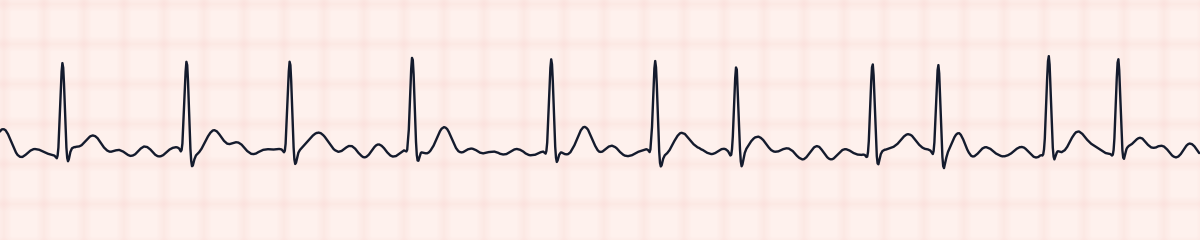
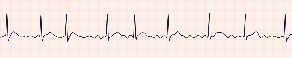
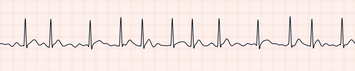
A rhythm is a set of rules, not a picture. These three strips all meet the rules for atrial fibrillation, with different numbers.
What atrial fibrillation gets mistaken for
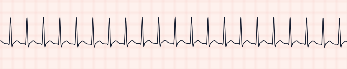
Atrial fibrillation vs. supraventricular tachycardia (SVT)
At fast rates the irregularity of AFib gets harder to see and it can pass for SVT. Put calipers on several R-R intervals: SVT is perfectly regular, AFib with a rapid ventricular response is not.
Atrial fibrillation vs. second-degree AV block, type I (Wenckebach)
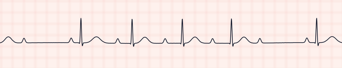
Wenckebach is also irregular, but it has clear P waves and its beats come in repeating groups. AFib has no P waves and no pattern at all.
Atrial fibrillation vs. ventricular fibrillation
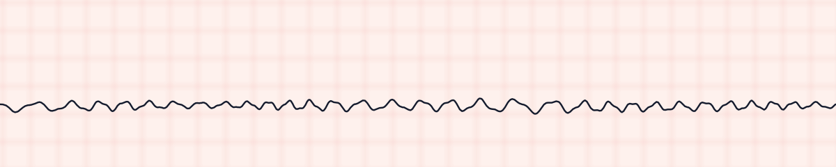
Both have a chaotic baseline. Atrial fibrillation still has distinct, narrow QRS complexes and the patient has a pulse. Ventricular fibrillation has no QRS complexes and no pulse.
Practice: atrial fibrillation or a look-alike?
Five strips. Some show atrial fibrillation; the others are the rhythms most often confused with it.
Strip 1 of 5
Lead II · 25 mm/s · 6 s
Causes of atrial fibrillation
- High blood pressure
- Heart failure and coronary artery disease
- Heart valve disease, especially of the mitral valve
- After cardiac surgery
- Overactive thyroid
- Alcohol, including binge drinking
- Sleep apnea and lung disease
- Increasing age
Why it matters
Atrial fibrillation is the most common sustained arrhythmia. It causes two problems. Blood that pools in the fibrillating atria can form clots that travel to the brain, so it is a major cause of stroke. And the loss of atrial contraction, together with a fast rate, lowers cardiac output and can bring on heart failure or low blood pressure.
What is done for atrial fibrillation
- Check whether the patient is stable: blood pressure, chest pain, shortness of breath, mental status.
- Count the apical pulse for a full minute. The radial pulse undercounts, because some beats are too weak to feel (a pulse deficit).
- Rate control with a beta blocker or diltiazem is the usual first step for a fast rate.
- Anticoagulation is considered to lower stroke risk.
- An unstable patient is treated with synchronized cardioversion.
- Report new-onset atrial fibrillation.
Study summary, not a protocol. Follow current American Heart Association guidelines and your facility’s policies. See the medical disclaimer.
Common mistakes with atrial fibrillation
- Calling coarse fibrillatory waves P waves. If the bumps do not have a fixed relationship to the QRS and the R-R is irregular, they are not P waves.
- Calculating the rate from one R-R interval. In an irregular rhythm only the 6-second count is valid.
- Missing AFib when the rate is fast because the R-R intervals look almost even.
Questions about atrial fibrillation
What does AFib look like on an EKG strip?
An irregularly irregular rhythm with no P waves. The baseline between QRS complexes is wavy, and the QRS complexes themselves are usually narrow and normal in shape.
How do you calculate the heart rate in atrial fibrillation?
Count the QRS complexes in a 6-second strip and multiply by 10. Methods that use the distance between two R waves do not work, because that distance keeps changing.
What is AFib with RVR?
RVR means rapid ventricular response: atrial fibrillation with a ventricular rate above 100 per minute. A rate of 100 or less is called controlled.
What is the difference between AFib and atrial flutter?
Atrial flutter has organized, sawtooth flutter waves at about 300 per minute and a ventricular rhythm that is often regular. Atrial fibrillation has a disorganized baseline and an irregular ventricular rhythm.