Ventricular rhythms
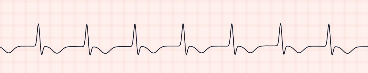
Accelerated idioventricular rhythm (AIVR) EKG strip
Accelerated idioventricular rhythm (AIVR) is a ventricular rhythm at 40 to 100 beats per minute. On an EKG strip it shows wide QRS complexes (0.12 seconds or more), regular, with no P waves in front of them. It is faster than a ventricular escape rhythm and slower than ventricular tachycardia.
Criteria from the American Heart Association guidelines and published ECG standards · Every strip checked by measurement · Sources and method · Updated November 15, 2026
- Rate
- 40–100
- PR interval
- None
- QRS
- ≥ 0.12 s
How to identify AIVR in five steps
Ask the same five questions of every strip. The right-hand column is what each one finds on the strip above.
1Rate
40 to 100. Some references give 50 to 110.
61 bpm
2Rhythm
Regular.
Regular
3P waves
None before the QRS. Unrelated sinus P waves may occasionally be seen.
None
4PR interval
None.
None
5QRS
0.12 s or wider, with a T wave pointing the opposite way.
0.16 s, wide
What is happening in the heart
A group of cells in the ventricle begins firing faster than its natural 20 to 40, fast enough to overtake the SA node for a while. This most often happens in the minutes after blood flow returns to heart muscle that was starved of it, which is why AIVR is known as a reperfusion rhythm. It typically starts gradually, runs for seconds to minutes, and stops by itself when the sinus rate overtakes it again.
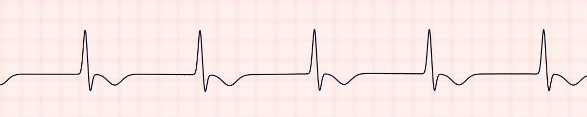
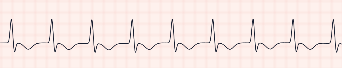
More AIVR strips: no two are the same
A rhythm is a set of rules, not a picture. These three strips all meet the rules for AIVR, with different numbers.
What AIVR gets mistaken for
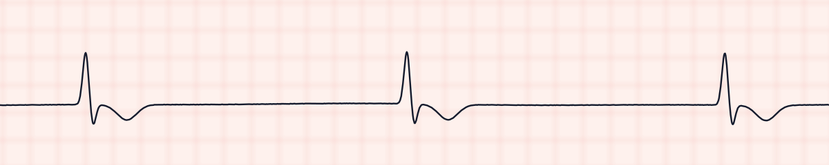
AIVR vs. idioventricular rhythm
The same wide complexes at 20 to 40 are an idioventricular escape rhythm, which is a failing heart’s last backup. AIVR is faster and is usually seen in a patient who is doing well.
AIVR vs. ventricular tachycardia
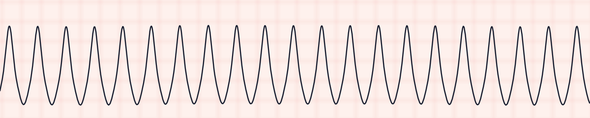
Ventricular tachycardia is the same shape at more than 100. Count the rate carefully before calling a wide rhythm V-tach: a wide rhythm at 80 is AIVR.
AIVR vs. ventricular paced rhythm
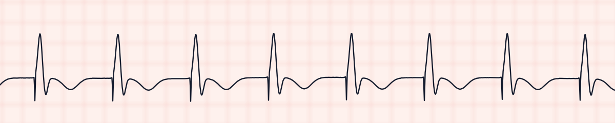
A ventricular paced rhythm is also wide, regular and in this range. Look at the very start of each QRS for a pacing spike.
Practice: AIVR or a look-alike?
Five strips. Some show AIVR; the others are the rhythms most often confused with it.
Strip 1 of 5
Lead II · 25 mm/s · 6 s
Causes of AIVR
- Reperfusion after a heart attack: thrombolytic drugs or an opened artery in the cath lab
- The first day or two after a myocardial infarction
- Digoxin toxicity
- Cardiomyopathy
- Return of circulation after a cardiac arrest
Why it matters
AIVR is the wide rhythm that usually should be left alone. After reperfusion it is often a sign that the artery has opened. Because its rate is near normal, cardiac output is generally maintained and the patient is comfortable. The danger lies in mistaking it for ventricular tachycardia and treating it.
What is done for AIVR
- Check the patient and the blood pressure. Most are stable.
- Count the rate to confirm it is under 100.
- Observe: it normally ends without treatment within minutes.
- Do not give antiarrhythmics to suppress it; they can remove a rhythm that is supporting the patient.
- If the patient is compromised, which is uncommon, raising the sinus rate with atropine may be considered.
- Report it, with the context, such as recent thrombolysis.
Study summary, not a protocol. Follow current American Heart Association guidelines and your facility’s policies. See the medical disclaimer.
Common mistakes with AIVR
- Calling it V-tach because it is wide, and reaching for amiodarone or the defibrillator.
- Not counting the rate.
- Missing a pacing spike and labeling a paced rhythm AIVR.
Questions about AIVR
What is the rate of AIVR?
Between 40 and 100 beats per minute, with some references using 50 to 110. Below that range it is an idioventricular rhythm; above it, ventricular tachycardia.
Is AIVR dangerous?
Usually not. It is typically brief, the patient is stable, and after treatment for a heart attack it is often a sign that blood flow has been restored. It rarely needs treatment.
How do you tell AIVR from V-tach?
By the rate. Both are wide and regular with no P waves. AIVR is 100 or less; ventricular tachycardia is over 100, usually 150 to 250.
Why is AIVR called a reperfusion arrhythmia?
Because it commonly appears just after blood flow is restored to heart muscle, for example after clot-dissolving drugs or angioplasty for a heart attack.