AV blocks
First-Degree AV Block With PVCs on an EKG Strip
Sinus rhythm with first-degree AV block and PVCs combines a conduction delay with an ectopic beat. Every sinus beat has a PR interval longer than 0.20 seconds, which is the first-degree block, and the sinus rhythm is interrupted by premature ventricular contractions, which come early, are wide and have no P wave ahead of them.
Criteria from the American Heart Association guidelines and published ECG standards · Every strip checked by measurement · Sources and method · Updated November 15, 2026
- Rate
- Underlying rhythm
- PR interval
- > 0.20 s on the sinus beats
- QRS
- Narrow, with wide PVCs
How to identify first-degree AV block with PVCs in five steps
Ask the same five questions of every strip. The right-hand column is what each one finds on the strip above.
1Rate
Count every QRS in six seconds and multiply by ten; the sinus rate underneath is usually 60 to 100.
~60 bpm
2Rhythm
Regular, except where a PVC comes early and a pause follows it.
Regular, with early beats
3P waves
One ahead of each sinus QRS; none ahead of a PVC.
None on the early beat
4PR interval
Over 0.20 seconds and identical on every sinus beat.
0.25 s on sinus beats, long
5QRS
Narrow on the sinus beats, 0.12 seconds or wider on the PVCs.
0.08 s; early beat 0.17 s
What is happening in the heart
The long PR interval comes from the AV node, which is passing every impulse on but taking longer than normal to do it. The PVCs come from somewhere else entirely: a spot in the ventricular muscle that fires before the next sinus beat is due. The sinus node is not disturbed by this and keeps to its schedule, so the P wave that falls during the PVC is hidden inside it and finds the ventricles unable to respond. The sinus beat after that arrives exactly on time, with the same long PR interval as before.
More first-degree AV block with PVCs strips: no two are the same
A rhythm is a set of rules, not a picture. These three strips all meet the rules for first-degree AV block with PVCs, with different numbers.
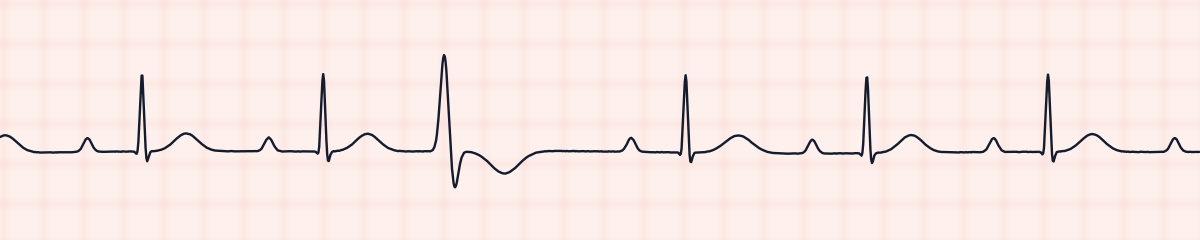
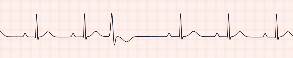
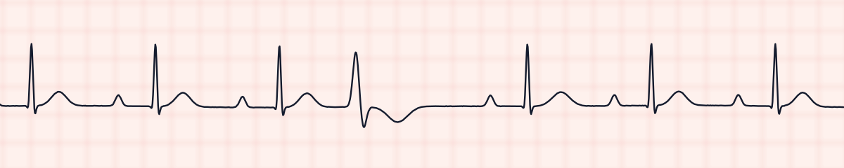
What first-degree AV block with PVCs get mistaken for
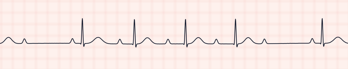
First-degree AV block with PVCs vs. second-degree AV block, type I (Wenckebach)
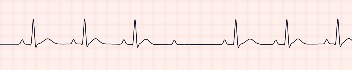
A pause plus a long PR interval suggests Wenckebach, but there the PR interval grows from beat to beat and the pause holds a P wave with no QRS. Here the PR is identical on every sinus beat and the pause comes after a wide early beat.
First-degree AV block with PVCs vs. second-degree AV block, type II (Mobitz II)
Mobitz II has a constant PR interval and a sudden pause too. Its pause contains a lone P wave; this one is preceded by a PVC.
First-degree AV block with PVCs vs. premature ventricular contractions (PVCs)
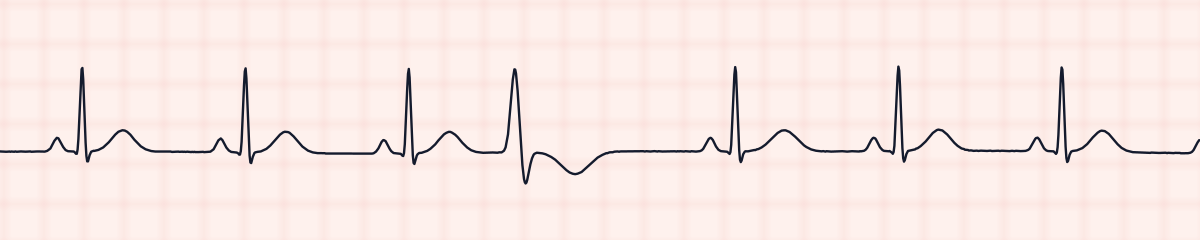
Sinus rhythm with PVCs has a normal PR interval. Measuring the PR on the sinus beats is the only way to find the first-degree block.
First-degree AV block with PVCs vs. first-degree AV block
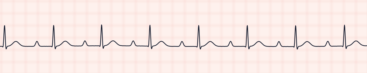
First-degree block by itself is perfectly regular, with no early beats.
Practice: first-degree AV block with PVCs or a look-alike?
Five strips. Some show first-degree AV block with PVCs; the others are the rhythms most often confused with it.
Strip 1 of 5
Lead II · 25 mm/s · 6 s
Causes of first-degree AV block with PVCs
- Coronary artery disease, including an inferior heart attack, which can affect both the AV node and the ventricular muscle
- Digoxin, which slows AV conduction and in excess provokes PVCs
- Low potassium or magnesium
- A beta blocker or calcium channel blocker (lengthening the PR) alongside any cause of PVCs
- Cardiomyopathy or heart failure
- An ageing conduction system in a person who also has benign PVCs
Why it matters
Either finding alone is usually well tolerated. Together they prompt a closer look for a shared cause, and digoxin toxicity heads that list because it produces exactly this pairing. The combination is also a reminder to watch the trend: a PR interval that keeps lengthening, dropped beats, or PVCs that become frequent or consecutive are all changes to report.
What is done for first-degree AV block with PVCs
- Measure the PR interval on a sinus beat away from the PVCs and document it.
- Count the PVCs and note whether they share one shape.
- Review medications that slow the AV node, and find out whether the patient takes digoxin.
- Expect potassium and magnesium results to be requested.
- Continue monitoring; report a lengthening PR interval, any non-conducted P wave, or increasing PVCs.
Study summary, not a protocol. Follow current American Heart Association guidelines and your facility’s policies. See the medical disclaimer.
Common mistakes with first-degree AV block with PVCs
- Finding the PVCs and stopping there. The eye goes to the wide beats and the long PR interval is missed.
- Measuring the PR interval only on the beat right after a PVC. Use several sinus beats.
- Reading the pause after a PVC as a dropped beat and calling the strip a second-degree block.
- Mistaking the T wave of the PVC for a P wave.
Questions about first-degree AV block with PVCs
How do you interpret a strip with more than one abnormality?
Work through the usual steps for the underlying rhythm: rate, regularity, P waves, PR interval, QRS. Then describe whatever interrupts it. The result here is sinus rhythm with first-degree AV block and PVCs.
Does a PVC change the PR interval?
Not in this rhythm. The sinus beats before and after the PVC have the same prolonged PR interval. The PVC itself has no PR interval because no P wave conducts to it.
Is first-degree AV block with PVCs serious?
Usually it is monitored, not treated. It needs prompt reporting if it is new in a patient taking digoxin, if the PVCs become frequent, or if beats start to be dropped.
Why is there a pause after the PVC?
The sinus impulse that arrives during the PVC cannot be conducted because the ventricles are still recovering. The next sinus beat comes at its normal time, which makes the gap after the PVC look long.