Sinus rhythms
Bundle Branch Block on an EKG Strip
A bundle branch block is a delay in one of the two branches that carry the impulse down through the ventricles. The rhythm itself is unchanged, most often a sinus rhythm with a P wave before every QRS and a normal PR interval, but each QRS lasts 0.12 seconds or longer and is often notched, because the two ventricles are no longer activated together.
Criteria from the American Heart Association guidelines and published ECG standards · Every strip checked by measurement · Sources and method · Updated November 15, 2026
- Rate
- 60–100
- PR interval
- 0.12–0.20 s
- QRS
- ≥ 0.12 s
How to identify a bundle branch block in five steps
Ask the same five questions of every strip. The right-hand column is what each one finds on the strip above.
1Rate
That of the underlying rhythm: 60 to 100 when it is a sinus rhythm.
96 bpm
2Rhythm
Regular, like the rhythm it rides on.
Regular
3P waves
Upright and uniform, each one followed by a QRS.
1 per QRS
4PR interval
0.12 to 0.20 seconds, with no change from beat to beat.
0.18 s
5QRS
0.12 seconds or longer on every beat, often notched like the letter M.
0.13 s, wide and notched
What is happening in the heart
Below the bundle of His the conduction system divides into a right and a left bundle branch. When one branch is blocked, the ventricle on that side is not reached by the fast route. It is activated late, by the impulse creeping across from the other ventricle through ordinary muscle. Two activations that should overlap now happen one after the other, which stretches the QRS and can give it two peaks. A single monitoring lead shows that a block is present; deciding whether it is the right or the left branch takes leads V1 and V6 of a 12-lead EKG.
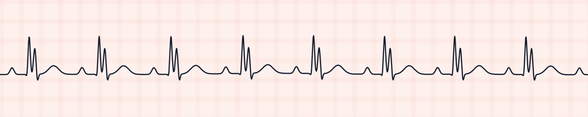
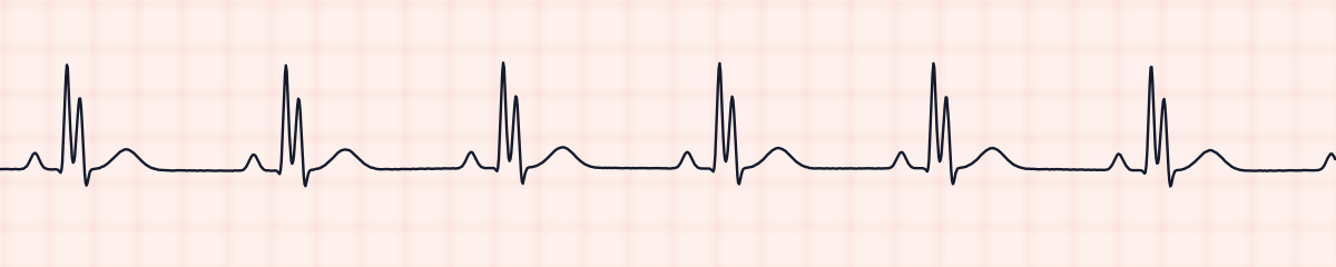
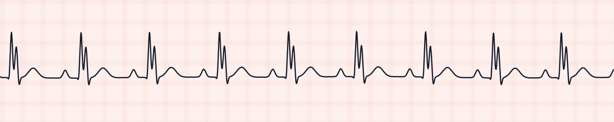
More bundle branch block strips: no two are the same
A rhythm is a set of rules, not a picture. These three strips all meet the rules for a bundle branch block, with different numbers.
What a bundle branch block gets mistaken for
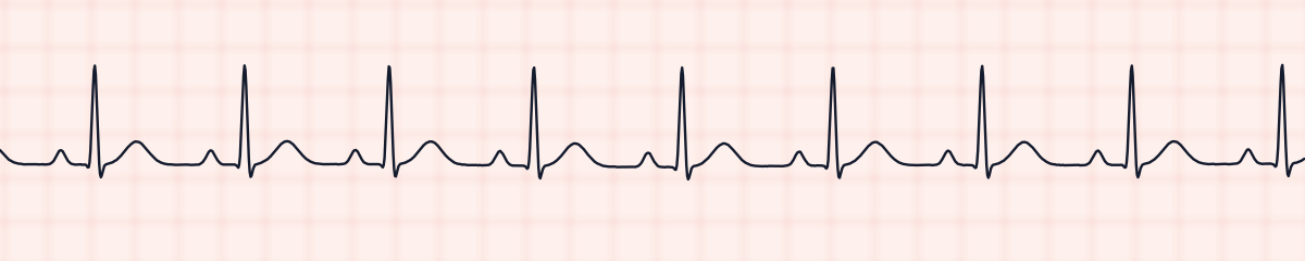
Bundle branch block vs. normal sinus rhythm
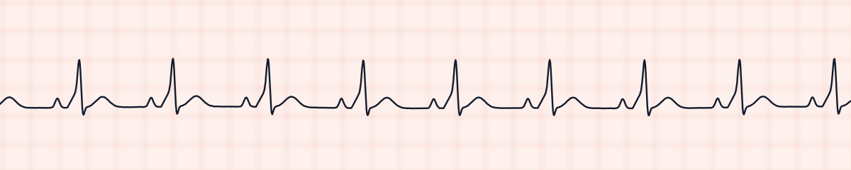
Everything matches normal sinus rhythm except the width of the QRS. Measure it: under 0.12 seconds is normal conduction, while 0.12 or more with this P wave relationship is a bundle branch block.
Bundle branch block vs. Wolff-Parkinson-White pattern (pre-excitation)
Both put a P wave in front of a wide QRS. In WPW the PR interval is short and the QRS begins with a slur; in a bundle branch block the PR is normal, the QRS begins crisply, and the extra width is at its end.
Bundle branch block vs. accelerated idioventricular rhythm (AIVR)
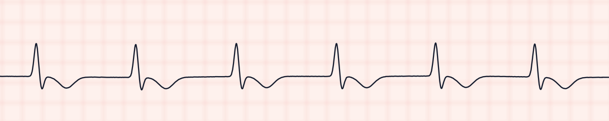
An accelerated idioventricular rhythm is also wide and regular at a similar rate, but no P wave is tied to each QRS. Look for a P wave at a fixed PR interval before calling any wide rhythm ventricular.
Practice: a bundle branch block or a look-alike?
Five strips. Some show a bundle branch block; the others are the rhythms most often confused with it.
Strip 1 of 5
Lead II · 25 mm/s · 6 s
Causes of a bundle branch block
- Coronary artery disease, including a heart attack
- Long-standing high blood pressure
- Cardiomyopathy and heart failure
- Aortic valve disease
- Age-related scarring of the conduction system
- Strain on the right heart, as in a pulmonary embolism or chronic lung disease (right bundle)
- A normal variant: some healthy people have a right bundle branch block
- A fast heart rate that outruns the recovery of one branch (a rate-related block)
Why it matters
An old, stable bundle branch block usually causes no symptoms and needs no treatment of its own. The question that matters is whether it is new. A new left bundle branch block in a patient with chest pain is handled as a possible heart attack until shown otherwise, and a left bundle branch block makes the ST segments of a 12-lead harder to interpret. It also tells you that part of the conduction system is already damaged, so an AV block appearing on top of it is taken seriously.
What is done for a bundle branch block
- Compare with earlier strips or EKGs to see whether the wide QRS is old or new.
- Document the QRS duration along with the underlying rhythm.
- Report a newly wide QRS, above all with chest pain, breathlessness, fainting or a fall in blood pressure.
- Expect a 12-lead EKG to identify which branch is blocked.
- No treatment is aimed at a chronic bundle branch block itself; care is directed at its cause.
Study summary, not a protocol. Follow current American Heart Association guidelines and your facility’s policies. See the medical disclaimer.
Common mistakes with a bundle branch block
- Calling a wide QRS ventricular without looking for P waves. A wide QRS that follows a P wave at a constant, normal PR interval started above the ventricles.
- Trying to name the right or left branch from a single monitoring lead.
- Reporting only “sinus rhythm” and leaving the QRS duration out.
- Assuming the block has always been there instead of checking the previous strip.
Questions about a bundle branch block
What does a bundle branch block look like on a rhythm strip?
A regular rhythm with a P wave before each QRS and a normal PR interval, in which every QRS is three small squares (0.12 seconds) or wider. The QRS is often notched, a shape sometimes called rabbit ears.
Can you tell right from left bundle branch block on a monitor strip?
Not reliably. The distinction depends on the shape of the QRS in leads V1 and V6. A rhythm strip in lead II tells you a block is present; a 12-lead EKG tells you which one.
Is a bundle branch block dangerous?
Often it is a long-standing finding without symptoms. It becomes urgent when it is new, particularly a new left bundle branch block with chest pain, or when it appears together with a second- or third-degree AV block.
How do you name the rhythm when there is a bundle branch block?
Name the underlying rhythm, then the conduction problem: for example, sinus rhythm with a bundle branch block, followed by the measured QRS duration.