Rhythm library
EKG rhythms, labeled and compared
Forty-two rhythms that nursing school, telemetry competency exams and ACLS expect you to recognize on a 6-second strip. They are grouped by where the rhythm comes from, because that is what decides how the strip looks. Each has its own page with measured examples and a short drill.
Jump to: Sinus rhythms · AV blocks · Supraventricular rhythms · Junctional rhythms · Ventricular rhythms · Paced rhythms · Arrest rhythms · Comparison chart
Sinus rhythms
The SA node is in charge and every beat has a normal P wave. They differ in rate and in how evenly the beats are spaced. Learn the normal numbers first.
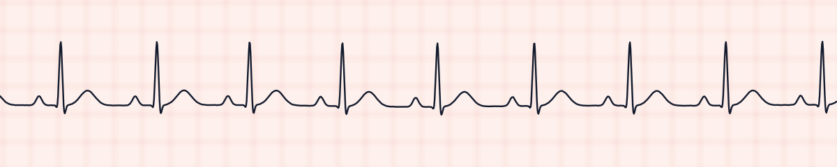
Normal sinus rhythm
Regular, with one P wave in front of every QRS. The yardstick for everything else.
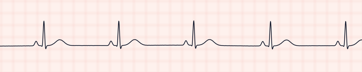
Sinus bradycardia
Normal sinus rhythm in every way except the rate, which is below 60.
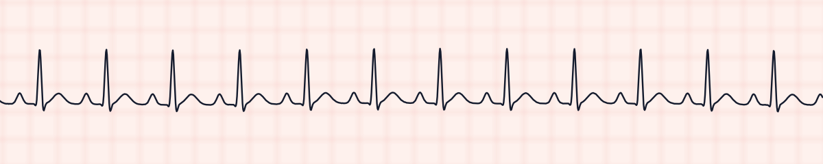
Sinus tachycardia
Normal sinus rhythm in every way except the rate, which is above 100. The P waves are still there.
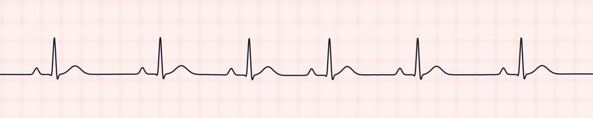
Sinus arrhythmia
Sinus rhythm that speeds up and slows down gradually. Every beat has a P wave and none is early.
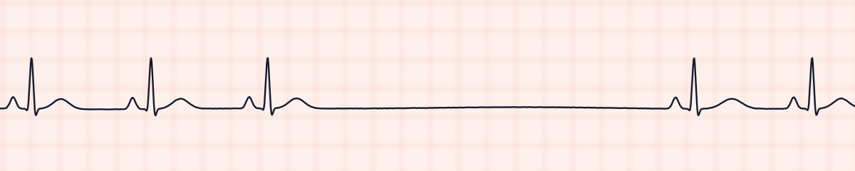
Sinus arrest (sinus pause)
Sinus rhythm interrupted by a pause with no P wave and no QRS.
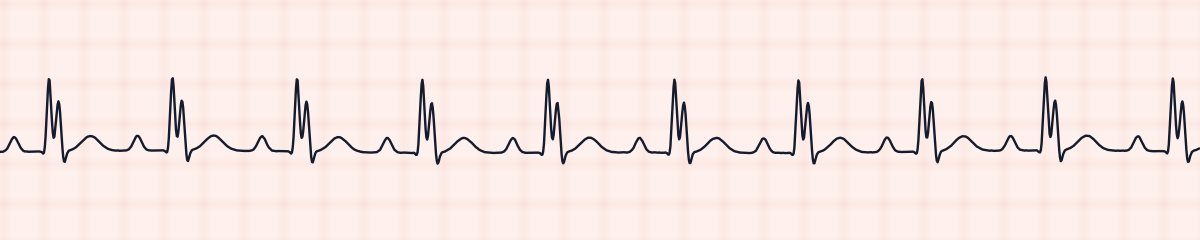
Sinus rhythm with bundle branch block
A P wave before every QRS and a normal PR interval, but the QRS is wide and notched.
AV blocks
The P waves are normal. What changes is whether, and how late, each one reaches the ventricles. Decided by the PR interval.
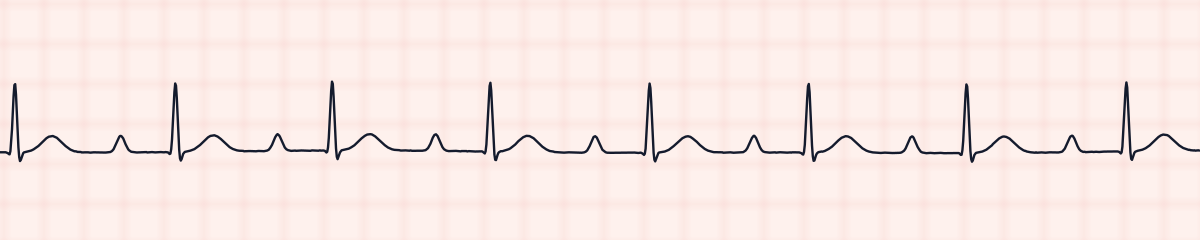
First-degree AV block
Every P wave conducts, but the PR interval is long and stays the same.
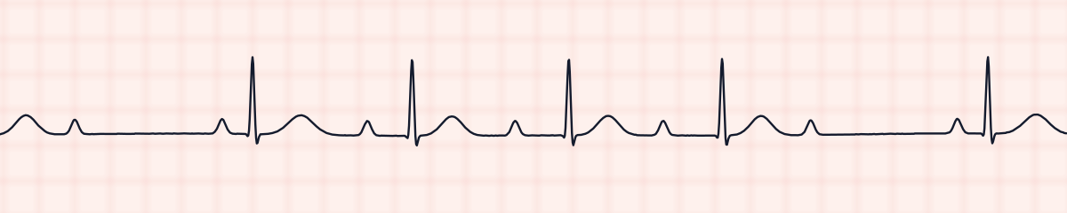
Second-degree AV block, type I (Wenckebach)
The PR interval gets longer, longer, longer, then a QRS is dropped.
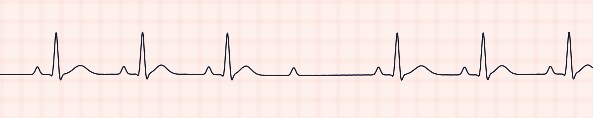
Second-degree AV block, type II (Mobitz II)
The PR interval never changes, then a QRS is dropped without warning.
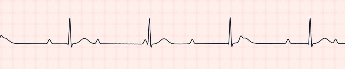
Third-degree (complete) AV block
P waves and QRS complexes are each regular, but have nothing to do with each other.
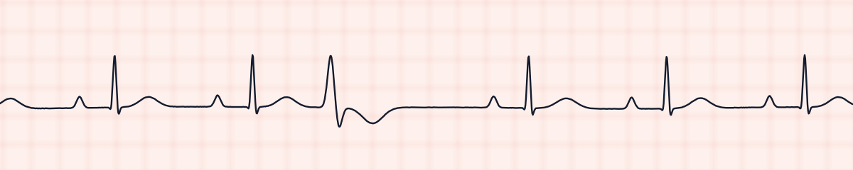
Sinus rhythm with first-degree AV block and PVCs
Two findings: a PR interval over 0.20 s on every sinus beat, and early wide beats with no P wave.
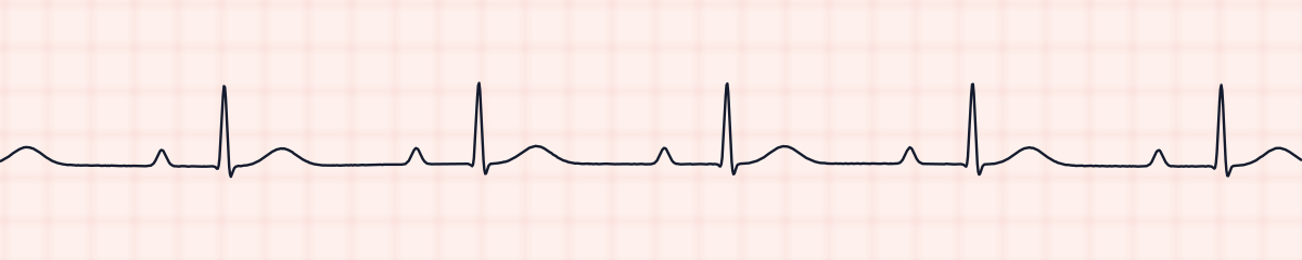
Sinus bradycardia with first-degree AV block
Two findings: a sinus rate under 60, and a PR interval over 0.20 s on every beat.
Supraventricular rhythms
They start in the atria, outside the SA node, so the QRS is narrow. Decided by regularity and by what the baseline looks like between complexes.
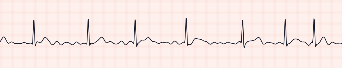
Atrial fibrillation
Irregularly irregular, with a wavy baseline and no P waves you can point to.
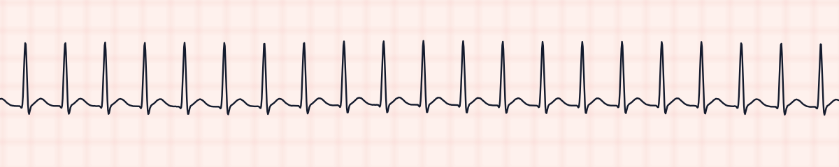
Supraventricular tachycardia (SVT)
Fast, narrow and perfectly regular. The P waves are buried.
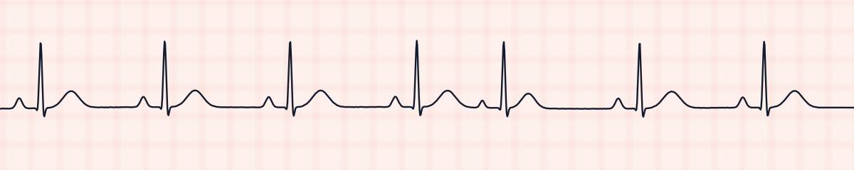
Premature atrial contractions (PACs)
A beat that comes early, with a P wave of a different shape and a normal narrow QRS.
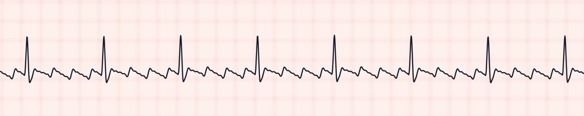
Atrial flutter
Sawtooth flutter waves at about 300 a minute, with a QRS after every third or fourth one.
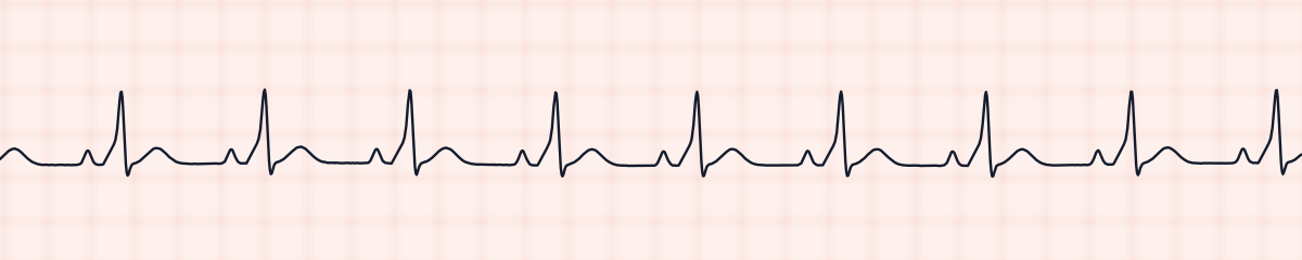
Wolff-Parkinson-White pattern (pre-excitation)
A short PR interval, then a slurred start to a wide QRS: the delta wave.
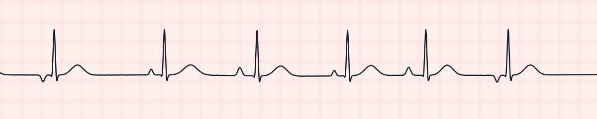
Wandering atrial pacemaker
P waves of at least three different shapes, at a rate under 100.
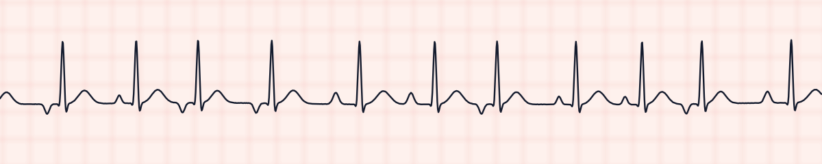
Multifocal atrial tachycardia (MAT)
Irregular and faster than 100, with P waves of at least three different shapes.
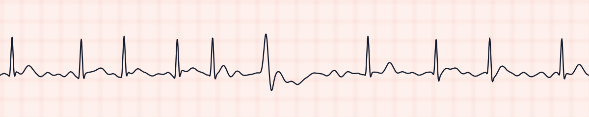
Atrial fibrillation with PVCs
Irregularly irregular with no P waves, plus a wide beat or two that looks nothing like the rest.
Junctional rhythms
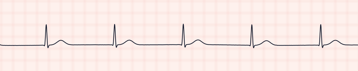
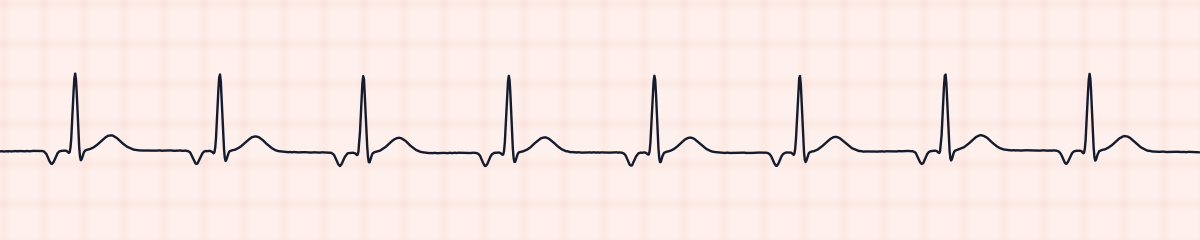
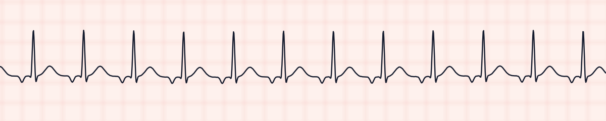
They start in the AV junction: a narrow QRS with no normal P wave in front of it. Decided by the rate.
Ventricular rhythms
They start in the ventricles, so the beat is wide and has no P wave. Decided by the rate, and by whether it is one beat or all of them.
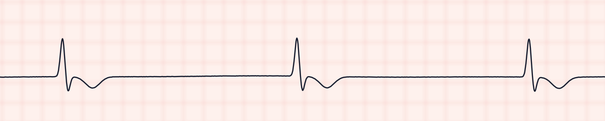
Idioventricular rhythm
Slow, wide and regular, with no P waves. The ventricles are pacing themselves.
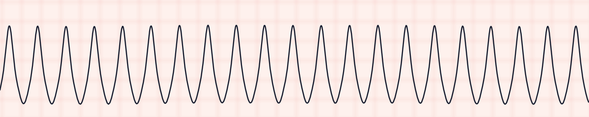
Ventricular tachycardia
Shockable if pulselessFast, wide and regular. Always check for a pulse.
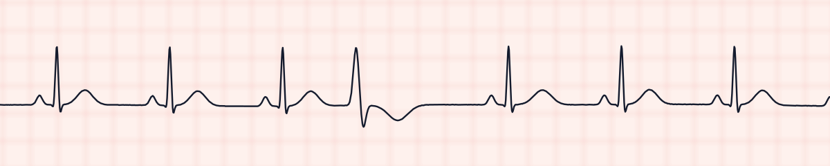
Premature ventricular contractions (PVCs)
A beat that comes early and is wide, with no P wave and a T wave pointing the other way.
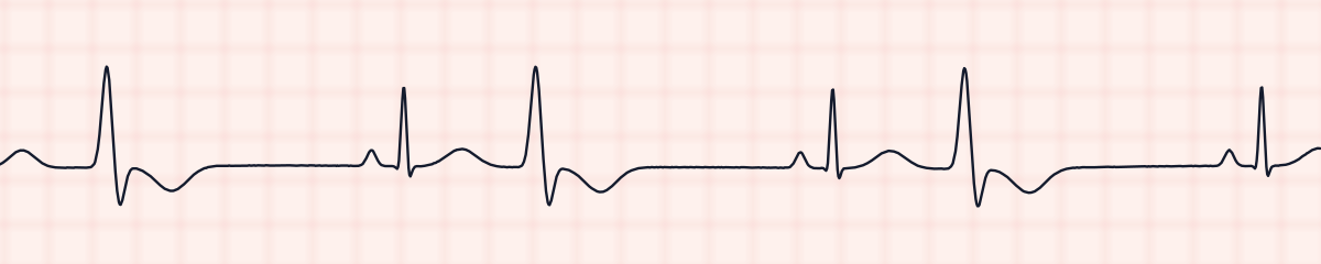
Ventricular bigeminy
Every other beat is a PVC: normal, wide, normal, wide.
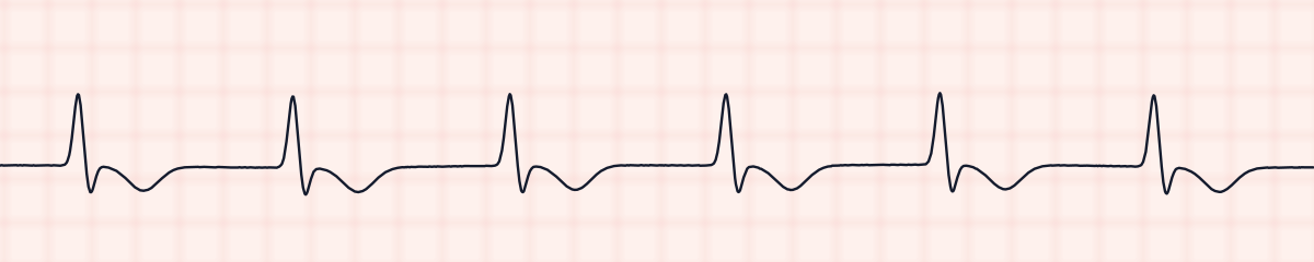
Accelerated idioventricular rhythm (AIVR)
Wide and regular with no P waves, at 40 to 100: too fast for an escape rhythm, too slow for V-tach.
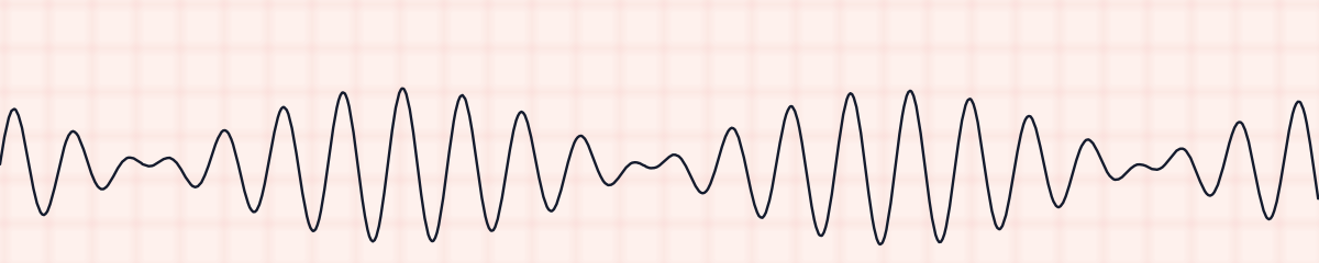
Torsades de pointes
A fast wide rhythm whose height swells and shrinks, as if twisting around the baseline.
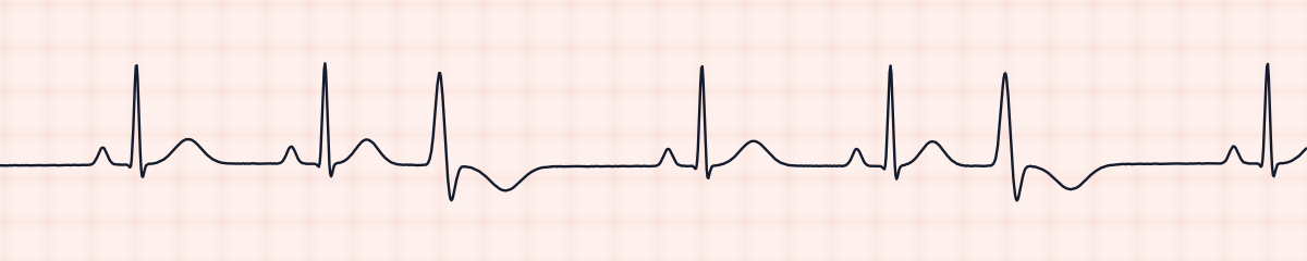
Ventricular trigeminy
Every third beat is a PVC: normal, normal, wide.
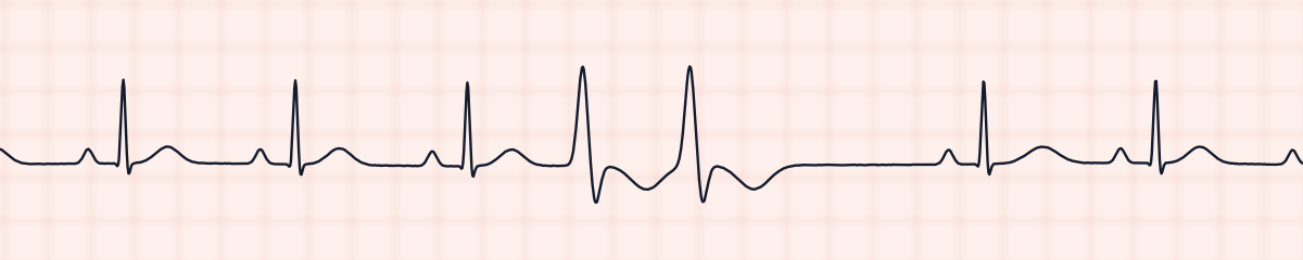
Ventricular couplet (paired PVCs)
Two PVCs back to back, with no normal beat between them.
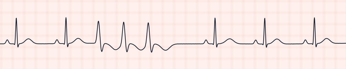
Ventricular triplet (three PVCs in a row)
Three PVCs in a row: the shortest run of ventricular tachycardia.
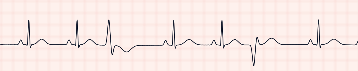
Multifocal PVCs
PVCs of more than one shape on the same strip, because they start in more than one place.
Paced rhythms
A pacemaker starts the beat. The pacing spike is the thing to find.
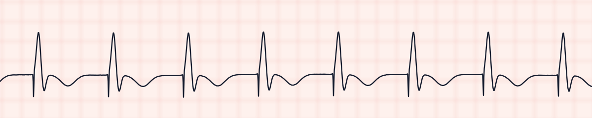
Ventricular paced rhythm
A sharp pacing spike, then a wide QRS, at a fixed rate.
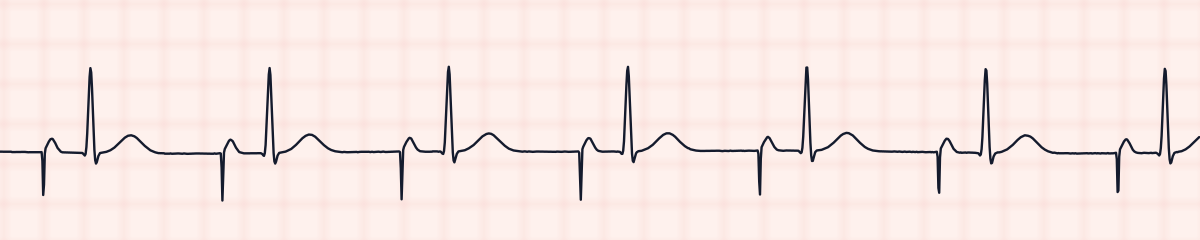
Atrial paced rhythm
A pacing spike, then a P wave, then a normal narrow QRS.
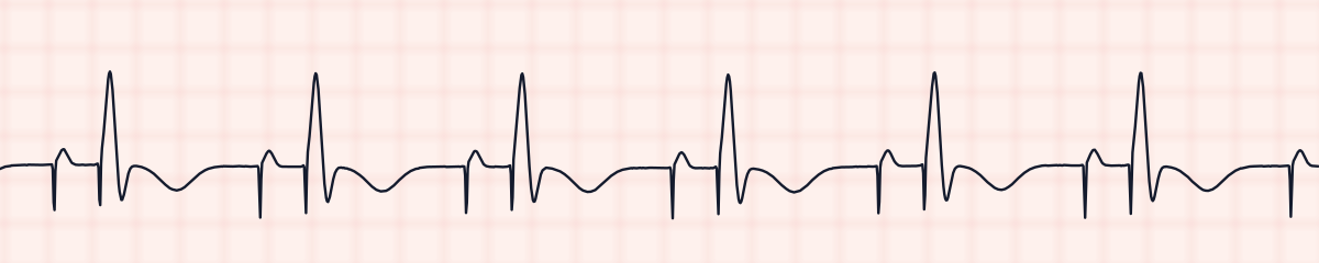
AV sequential paced rhythm
Two pacing spikes per beat: one before the P wave and one before a wide QRS.
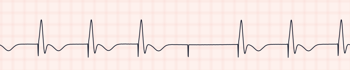
Pacemaker failure to capture
A pacing spike with no QRS after it.
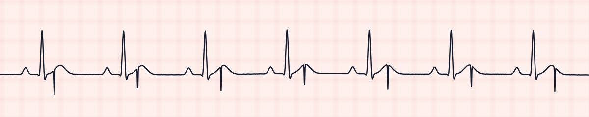
Pacemaker failure to sense
Pacing spikes that land on the patient's own beats, where no pacing was needed.
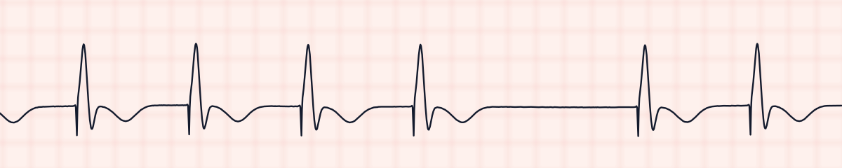
Pacemaker failure to pace (failure to fire)
A paced rhythm with a pause in which there is no spike and no QRS.
Arrest rhythms
The patient has no pulse. Two are shockable and two are not, and that is the first thing to know about each.
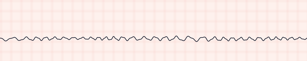
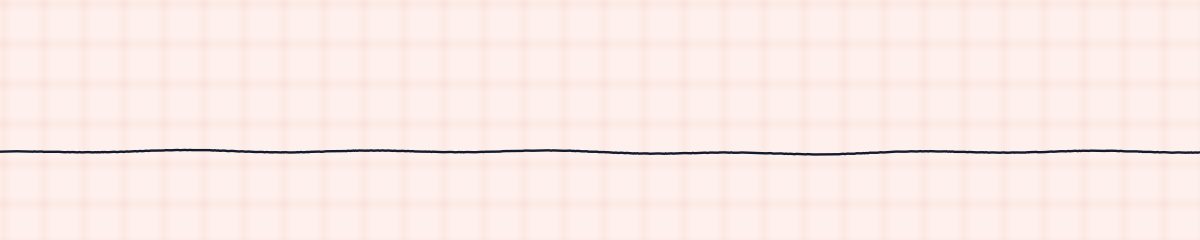
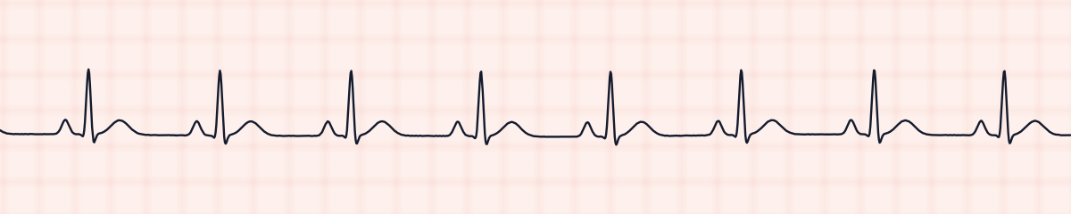
EKG rhythms comparison chart
The five-step read for every rhythm, side by side. Read across a row to describe a rhythm; read down a column to see which step separates two rhythms that look alike.
| Rhythm | Rate | Regularity | P waves | PR interval | QRS |
|---|---|---|---|---|---|
| Normal sinus rhythm | 60–100 | Regular | 1 per QRS | 0.12–0.20 s | Narrow |
| First-degree AV block | Usually 60–100 | Regular | 1 per QRS | > 0.20 s, constant | Narrow |
| Second-degree type I (Wenckebach) | Atrial > ventricular | Grouped | More Ps than QRSs | Lengthens, then a drop | Narrow |
| Second-degree type II (Mobitz II) | Atrial > ventricular | Irregular at the drop | More Ps than QRSs | Constant, then a drop | Often wide |
| Third-degree AV block | Ventricular 20–60 | Regular | Regular, unrelated to QRS | Never repeats | Narrow or wide |
| Atrial fibrillation | Variable | Irregularly irregular | None, wavy baseline | None | Narrow |
| SVT | 150–250 | Regular | Hidden | None | Narrow |
| Idioventricular rhythm | 20–40 | Regular | None | None | Wide |
| Ventricular tachycardia | Over 100 | Regular | None | None | Wide |
| Ventricular fibrillation | Not countable | Chaotic | None | None | None |
| Asystole | 0 | None | None | None | None |
| PEA | Any | Organized | Any | Any | Any |
| Sinus bradycardia | Under 60 | Regular | 1 per QRS | 0.12–0.20 s | Narrow |
| Sinus tachycardia | 101–150 | Regular | 1 per QRS | 0.12–0.20 s | Narrow |
| Sinus arrhythmia | 60–100, varying | Irregular, gradual | 1 per QRS | 0.12–0.20 s | Narrow |
| Sinus arrest (pause) | Underlying sinus rate | Regular with a pause | 1 per QRS, none in the pause | 0.12–0.20 s | Narrow |
| Sinus rhythm with PACs | Underlying rhythm | Regular with an early beat | Early one looks different | Normal | Narrow |
| Atrial flutter | Atrial 250–350 | Regular or variable | Sawtooth flutter waves | None | Narrow |
| Junctional rhythm | 40–60 | Regular | Absent or inverted | None or < 0.12 s | Narrow |
| Accelerated junctional rhythm | 60–100 | Regular | Absent or inverted | None or < 0.12 s | Narrow |
| Sinus rhythm with PVCs | Underlying rhythm | Regular with an early beat | None on the early beat | Normal on sinus beats | Early beat wide |
| Ventricular bigeminy | Count every QRS | Pairs: short, long | On every other beat | Normal on sinus beats | Alternates narrow, wide |
| Accelerated idioventricular (AIVR) | 40–100 | Regular | None | None | Wide |
| Torsades de pointes | 200–250 | Fast, twisting | None | None | Wide, changing |
| Paced rhythm | Set rate, 60–80 | Regular | None | None; spike before QRS | Wide |
| Junctional tachycardia | Over 100 | Regular | Absent or inverted | None or < 0.12 s | Narrow |
| Ventricular trigeminy | Count every QRS | Threes: normal, short, long | None on every third beat | Normal on sinus beats | Every third one wide |
| PVC couplet | Underlying rhythm | Regular with two early beats | None on the pair | Normal on sinus beats | Two wide in a row |
| PVC triplet (3-beat run of V-tach) | Underlying rhythm | Regular with a 3-beat run | None in the run | Normal on sinus beats | Three wide in a row |
| Multifocal PVCs | Underlying rhythm | Regular with early beats | None on the early beats | Normal on sinus beats | Wide, of different shapes |
| Atrial paced rhythm | Set rate, 60–80 | Regular | Each after a spike | Normal, spike to QRS | Narrow |
| AV paced rhythm | Set rate, 60–80 | Regular | Each after a spike | Fixed delay between two spikes | Wide |
| Failure to capture | Below the set rate | Regular with a missed beat | None | None; a spike with no QRS | Wide |
| Sinus rhythm with bundle branch block | 60–100 | Regular | 1 per QRS | 0.12–0.20 s | Wide, notched |
| Wolff-Parkinson-White (WPW) | 60–100 | Regular | 1 per QRS | < 0.12 s | Wide, with a delta wave |
| Wandering atrial pacemaker | Under 100 | Slightly irregular | 3 or more shapes | Changes with the P wave | Narrow |
| Multifocal atrial tachycardia (MAT) | Over 100 | Irregularly irregular | 3 or more shapes | Changes with the P wave | Narrow |
| Failure to sense | The patient’s own rate | Underlying rhythm | Those of the underlying rhythm | Normal; spikes fall after the QRS | Narrow |
| Failure to pace | Below the set rate | Regular with a pause | None | None; no spike in the pause | Wide |
| Atrial fibrillation with PVCs | Count every QRS | Irregularly irregular | None, wavy baseline | None | Narrow, with wide PVCs |
| First-degree AV block with PVCs | Underlying sinus rate | Regular with early beats | None on the early beats | > 0.20 s on sinus beats | Narrow, with wide PVCs |
| Sinus bradycardia with first-degree AV block | Under 60 | Regular | 1 per QRS | > 0.20 s, constant | Narrow |
Narrow means a QRS under 0.12 s (3 small squares); wide means 0.12 s or more. Rates are per minute. PEA is any organized rhythm in a patient with no pulse, so it has no pattern of its own.
Three questions that sort most strips
- Is the QRS narrow or wide? Narrow means the impulse came from above the ventricles. Wide, with no P waves, means it started in the ventricles.
- Is it regular? An irregular narrow rhythm with no P waves is atrial fibrillation. An irregular rhythm with P waves is usually a second-degree block.
- Is there one P wave for every QRS, with the same PR interval? If yes, it is a sinus rhythm or first-degree block. If not, compare the PR intervals to find which block it is.
Built for rhythm-strip exams
Every rhythm in this library can be named from a single lead II strip, which is what a dysrhythmia exam puts in front of you. Each one is put through the same checks before it appears here; the method is on the about and methods page.
Try today’s three free stripsWhere this comes from
The criteria and rules on this page are taken from published sources, and every strip is drawn to them and then measured by a separate program before it is shown. How the strips are made and checked
- American Heart Association. CPR and Emergency Cardiovascular Care guidelines: adult cardiac arrest, bradycardia and tachycardia algorithms. Which rhythms are shockable, and the first actions for the arrest rhythms, slow rhythms and fast rhythms.
- American Heart Association, American College of Cardiology and Heart Rhythm Society. Recommendations for the standardization and interpretation of the electrocardiogram, part I (Circulation, 2007). The paper every strip is drawn on: 25 mm per second and 10 mm per millivolt.
- University of Arkansas. How to Interpret Rhythm Strips, in the open EKG Interpretation manual. The five-step read and the normal ranges for rate, PR interval and QRS.