AV blocks
Third-degree (complete) heart block EKG strip
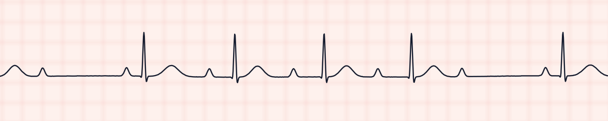
Third-degree heart block, or complete heart block, is the total failure of conduction between the atria and the ventricles. On an EKG strip the P waves are regular and the QRS complexes are regular, but each at its own rate and with no relationship to the other. The PR interval is different on every beat.
Criteria from the American Heart Association guidelines and published ECG standards · Every strip checked by measurement · Sources and method · Updated November 15, 2026
- Rate
- Ventricular 20–60
- PR interval
- Varies, no relationship
- QRS
- Narrow or wide
How to identify third-degree heart block in five steps
Ask the same five questions of every strip. The right-hand column is what each one finds on the strip above.
1Rate
Two rates. The atrial rate is the sinus rate, often 60 to 100. The ventricular rate is slower: 40 to 60 with a narrow QRS, 20 to 40 with a wide one.
43 bpm
2Rhythm
The P-P intervals are regular and the R-R intervals are regular. They are simply not the same interval.
Regular
3P waves
Normal, and more of them than QRS complexes. Some are hidden inside a QRS or a T wave.
72/min, unrelated
4PR interval
No true PR interval. The distance from P to QRS changes on every beat.
Varies
5QRS
Narrow if the escape rhythm is junctional, 0.12 s or wider if it is ventricular.
0.09 s
What is happening in the heart
No atrial impulse reaches the ventricles. The atria keep following the SA node, and a backup pacemaker below the block takes over the ventricles. If that pacemaker is in the AV junction, the QRS is narrow at about 40 to 60 per minute. If it is in the ventricles, the QRS is wide at about 20 to 40. The two rhythms run side by side, which is called AV dissociation.
More third-degree heart block strips: no two are the same
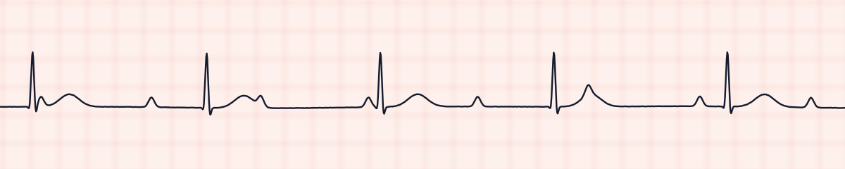
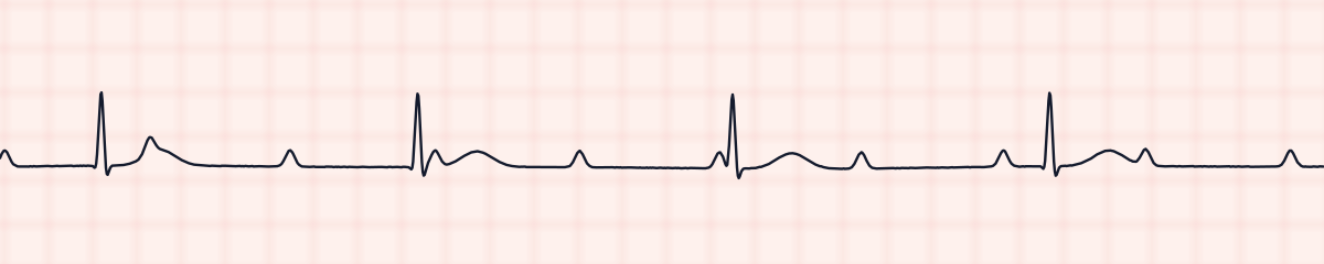
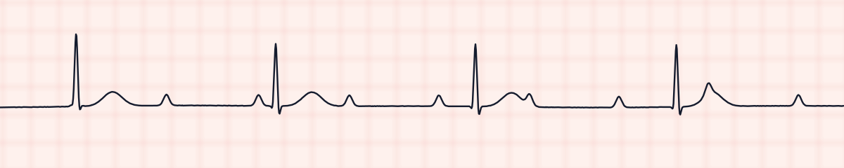
A rhythm is a set of rules, not a picture. These three strips all meet the rules for third-degree heart block, with different numbers.
What third-degree heart block gets mistaken for
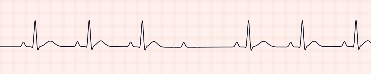
Third-degree heart block vs. second-degree AV block, type II (Mobitz II)
Mobitz II also has P waves without a QRS, but the beats that do conduct share one fixed PR interval, and the rhythm is irregular at the drop. In complete block no PR repeats and the R-R is regular.
Third-degree heart block vs. idioventricular rhythm
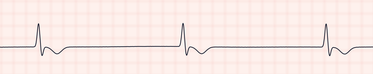
An idioventricular rhythm is the same slow, wide ventricular rhythm with no P waves at all. If you can march out regular P waves through the strip, it is complete heart block.
Third-degree heart block vs. second-degree AV block, type I (Wenckebach)
Both show a changing PR interval. In Wenckebach it changes in order, longer each beat, and the rhythm is irregular. In complete block it changes at random while the R-R stays regular.
Practice: third-degree heart block or a look-alike?
Five strips. Some show third-degree heart block; the others are the rhythms most often confused with it.
Strip 1 of 5
Lead II · 25 mm/s · 6 s
Causes of third-degree heart block
- Myocardial infarction, inferior or anterior
- Degeneration of the conduction system with age
- Drug toxicity: digoxin, beta blockers, calcium channel blockers
- After cardiac surgery or ablation
- Lyme disease and other infections of the heart
- Present from birth in some patients
Why it matters
The ventricles are depending on a backup pacemaker that is slow and can fail. Patients may faint, become confused or hypotensive, or arrest. A wide, slow escape rhythm is the least reliable. Complete heart block is treated as urgent even in a patient who looks comfortable.
What is done for third-degree heart block
- Assess the patient at once: level of consciousness, blood pressure, perfusion.
- Call for help and prepare transcutaneous pacing.
- Atropine may be tried but often fails, especially when the QRS is wide.
- Dopamine or epinephrine infusions support the rate until pacing is established.
- A permanent pacemaker is the definitive treatment in most cases.
Study summary, not a protocol. Follow current American Heart Association guidelines and your facility’s policies. See the medical disclaimer.
Common mistakes with third-degree heart block
- Reading it as first-degree block because one P wave happens to sit a long way in front of a QRS. Check several beats: the distance will not repeat.
- Counting only the P waves that are easy to see. Use calipers to march out the P-P interval and find the ones buried in QRS complexes and T waves.
- Calling it irregular. Both the atria and the ventricles are regular; they are out of step with each other.
Questions about third-degree heart block
How do you identify third-degree heart block on a strip?
Show three things: the P waves are regular, the QRS complexes are regular, and the PR interval is different on every beat. There are more P waves than QRS complexes, and the ventricular rate is slow.
What is AV dissociation?
AV dissociation means the atria and ventricles are beating independently, each driven by its own pacemaker. Complete heart block is the most common cause of it.
Is third-degree heart block an emergency?
It is treated as one. The ventricles rely on a slow escape rhythm that can stop. Symptomatic patients need pacing, and most patients need a permanent pacemaker.
Is the QRS wide or narrow in complete heart block?
Either. A junctional escape rhythm gives a narrow QRS at about 40 to 60 per minute. A ventricular escape rhythm gives a wide QRS at about 20 to 40 per minute.