Arrest rhythms
Asystole EKG strip
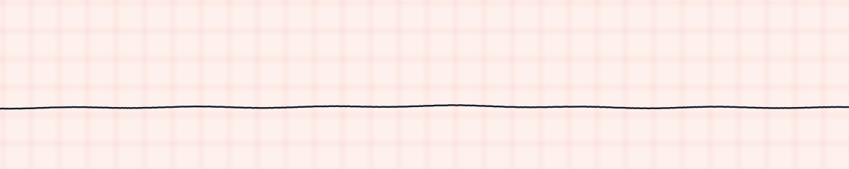
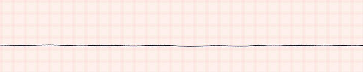
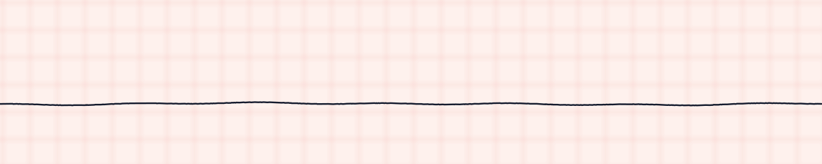
Asystole is the absence of electrical activity in the heart. On an EKG strip it is a flat or nearly flat line: no P waves, no QRS complexes and no T waves. It is a cardiac arrest rhythm, commonly called flatline, and it is not shockable.
Criteria from the American Heart Association guidelines and published ECG standards · Every strip checked by measurement · Sources and method · Updated November 15, 2026
- Rate
- 0
- PR interval
- None
- QRS
- None
How to identify asystole in five steps
Ask the same five questions of every strip. The right-hand column is what each one finds on the strip above.
1Rate
Zero.
0
2Rhythm
None.
None
3P waves
None. If P waves continue with no QRS complexes, it is called ventricular standstill and is treated the same way.
None
4PR interval
None.
None
5QRS
None.
None
What is happening in the heart
No pacemaker in the heart is firing, so there is nothing to conduct and nothing to contract. It is usually the end stage of another problem: ventricular fibrillation that has burned itself out, a slow rhythm that has kept slowing, or a long period without oxygen. Because there is no disorganized electrical activity to reset, a shock has nothing to work on.
More asystole strips: no two are the same
A rhythm is a set of rules, not a picture. These three strips all meet the rules for asystole, with different numbers.
What asystole gets mistaken for
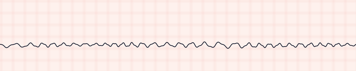
Asystole vs. ventricular fibrillation
Fine ventricular fibrillation is a low, irregular wobble that does not stop. Increase the gain and look in a second lead. If the movement is real and persistent, treat it as VF and shock it.
Asystole vs. pulseless electrical activity (PEA)
Both are non-shockable arrest rhythms with the same treatment. The monitor is what differs: asystole is a flat line, and PEA shows organized complexes.
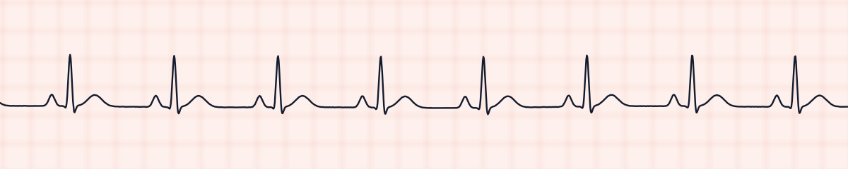
Practice: asystole or a look-alike?
Five strips. Some show asystole; the others are the rhythms most often confused with it.
Strip 1 of 5
Lead II · 25 mm/s · 6 s
Causes of asystole
- Prolonged ventricular fibrillation or pulseless electrical activity
- Severe, prolonged hypoxia
- Very high potassium
- Severe acidosis
- Hypothermia
- Drug overdose
- Massive heart attack or pulmonary embolism
Why it matters
Asystole has the poorest outlook of the arrest rhythms, because it usually means the heart has been without oxygen for a long time. The one chance is to find a cause that can be reversed. It is also the rhythm that is most often a false alarm: a lead that has fallen off draws the same flat line.
What is done for asystole
- Check the patient first. A flat line in someone who is awake is an equipment problem.
- Confirm true asystole: check that the leads are attached, look in a second lead, and turn up the gain.
- Start CPR immediately.
- Give epinephrine during the resuscitation.
- Do not defibrillate. Asystole is not a shockable rhythm.
- Search for reversible causes, the Hs and Ts.
Study summary, not a protocol. Follow current American Heart Association guidelines and your facility’s policies. See the medical disclaimer.
Common mistakes with asystole
- Shocking asystole. A shock does not start a heart that has no electrical activity.
- Starting a resuscitation on a disconnected lead, or the opposite: assuming a flat line is a loose lead without looking at the patient.
- Missing fine VF because the gain was set low.
Questions about asystole
What does asystole look like on an EKG strip?
A flat or almost flat line with no waves or complexes. A slight slow drift of the baseline is normal; there are no P waves, QRS complexes or T waves.
Is asystole a shockable rhythm?
No. Defibrillation works by stopping disorganized electrical activity so that a normal pacemaker can restart. In asystole there is no activity to stop. Treatment is CPR, epinephrine and fixing the cause.
Why do you check asystole in two leads?
To make sure it is real. Fine ventricular fibrillation can look flat in one lead and visible in another, and a loose or disconnected lead also produces a flat line.