Paced rhythms
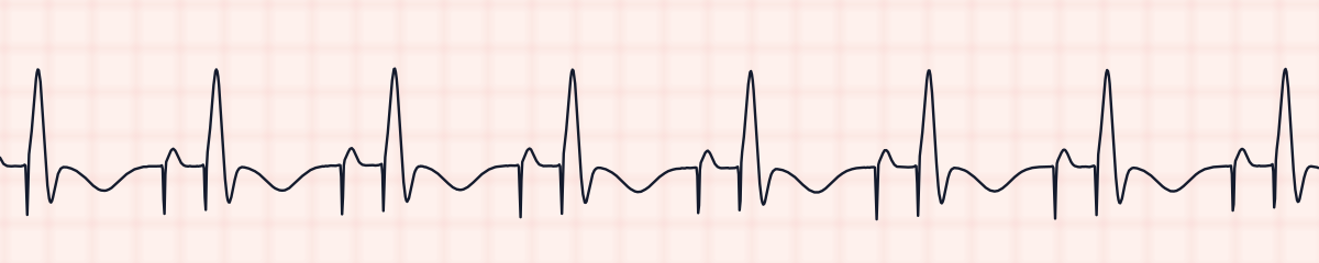
AV sequential paced rhythm EKG strip
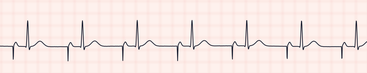
An AV sequential paced rhythm, or dual-chamber pacing, is produced by a pacemaker that stimulates the atria and then the ventricles. On an EKG strip each beat has two pacing spikes: the first is followed by a P wave, and the second, a set interval later, by a wide QRS.
Criteria from the American Heart Association guidelines and published ECG standards · Every strip checked by measurement · Sources and method · Updated November 15, 2026
- Rate
- Set rate, often 60–80
- PR interval
- The programmed AV delay
- QRS
- ≥ 0.12 s
How to identify an AV paced rhythm in five steps
Ask the same five questions of every strip. The right-hand column is what each one finds on the strip above.
1Rate
The set rate, commonly 60 to 80.
64 bpm
2Rhythm
Regular.
Regular
3P waves
One per beat, right after the first spike.
Each after a pacing spike
4PR interval
The gap between the two spikes is the programmed AV delay, usually 0.12 to 0.20 s, and never varies.
0.20 s between the spikes
5QRS
0.12 s or wider, beginning at the second spike.
0.15 s, wide
What is happening in the heart
The pacemaker has two leads, one in the right atrium and one in the right ventricle. It fires the atrial lead, waits for the programmed AV delay, which stands in for the PR interval, and then fires the ventricular lead. The ventricles are activated from the lead tip through muscle, so the QRS is wide. Pacing both chambers in order keeps the atria filling the ventricles just before they contract.
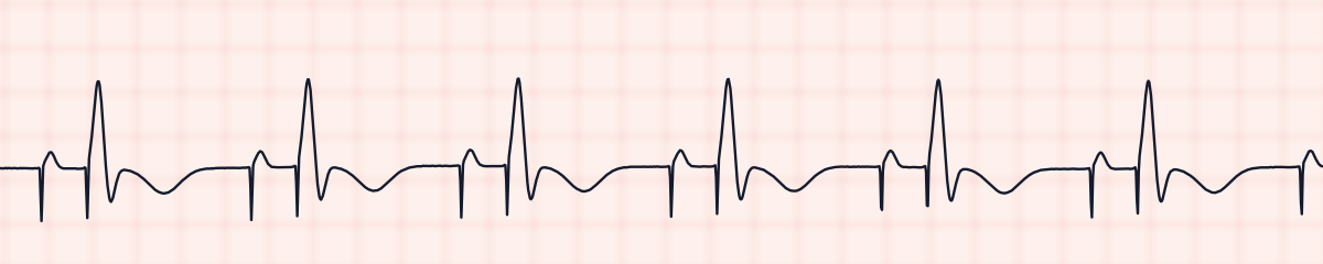
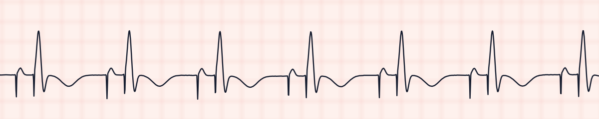
More AV paced rhythm strips: no two are the same
A rhythm is a set of rules, not a picture. These three strips all meet the rules for an AV paced rhythm, with different numbers.
What an AV paced rhythm gets mistaken for
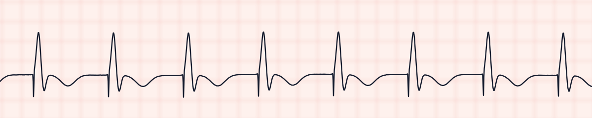
AV paced rhythm vs. ventricular paced rhythm
Ventricular pacing alone has one spike per beat, at the QRS, and no paced P wave. Look in front of each QRS for a second spike with a P wave behind it.
AV paced rhythm vs. atrial paced rhythm
Atrial pacing alone has one spike per beat, at the P wave, and a narrow QRS. A wide QRS that starts with its own spike means the ventricle is being paced as well.
Practice: an AV paced rhythm or a look-alike?
Five strips. Some show an AV paced rhythm; the others are the rhythms most often confused with it.
Strip 1 of 5
Lead II · 25 mm/s · 6 s
Why a patient is paced this way
- Complete heart block
- Second-degree AV block type II
- Sinus node disease together with AV node disease
- Temporary dual-chamber pacing after heart surgery
Why it matters
Dual-chamber pacing replaces the whole electrical sequence, so there are two things to verify on every beat. A dual-chamber device also adjusts to the patient: it may pace only the atria, only the ventricles, or both, depending on what the heart does by itself, so the number of spikes can change from beat to beat without anything being wrong.
What to check with an AV paced rhythm
- Confirm atrial capture (a P wave after the first spike) and ventricular capture (a QRS after the second).
- Check a pulse with the paced beats.
- Know the programmed lower rate.
- Report any spike that is not followed by its wave.
- For temporary pacing, check connections and settings as ordered.
Study summary, not a protocol. Follow current American Heart Association guidelines and your facility’s policies. See the medical disclaimer.
Common mistakes with an AV paced rhythm
- Counting both spikes as beats and doubling the heart rate.
- Assuming a malfunction when the device paces only one chamber for a while.
- Mistaking the atrial spike for artifact.
Questions about an AV paced rhythm
What does AV pacing look like on an EKG strip?
Two pacing spikes in each beat. The first is followed by a P wave, and the second by a wide QRS.
What is the AV delay?
The interval the pacemaker waits between pacing the atria and pacing the ventricles. On the strip it is the distance between the two spikes, and it takes the place of the PR interval.
Why does a dual-chamber pacemaker sometimes show only one spike?
It senses the heart’s own activity. If the atria fire by themselves it paces only the ventricles; if the ventricles respond by themselves it paces only the atria. That is normal function.