Ventricular rhythms
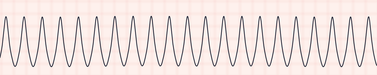
Idioventricular rhythm EKG strip
An idioventricular rhythm is a slow rhythm that starts in the ventricles when the pacemakers above them have failed. On an EKG strip it shows wide QRS complexes (0.12 seconds or more) at 20 to 40 beats per minute, regular, with no P waves. It is also called a ventricular escape rhythm.
Criteria from the American Heart Association guidelines and published ECG standards · Every strip checked by measurement · Sources and method · Updated November 15, 2026
- Rate
- 20–40
- PR interval
- None
- QRS
- ≥ 0.12 s
How to identify idioventricular rhythm in five steps
Ask the same five questions of every strip. The right-hand column is what each one finds on the strip above.
1Rate
20 to 40 per minute. From 40 to about 100 with the same shape it is called accelerated idioventricular rhythm (AIVR).
26 bpm
2Rhythm
Regular.
Regular
3P waves
None.
None
4PR interval
None.
None
5QRS
0.12 s or wider, with a T wave that points in the opposite direction.
0.16 s, wide
What is happening in the heart
The heart has backup pacemakers in order. When the SA node and the AV junction both fail to produce an impulse, cells in the ventricles begin to fire at their own slow built-in rate of 20 to 40 per minute. Because the impulse starts in the ventricular muscle and spreads cell to cell instead of through the fast conduction pathway, the QRS is wide and looks unlike a normal one, and the T wave points the opposite way.
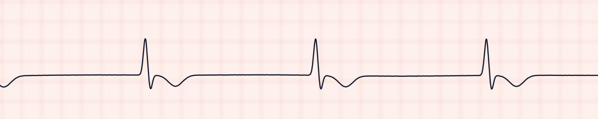
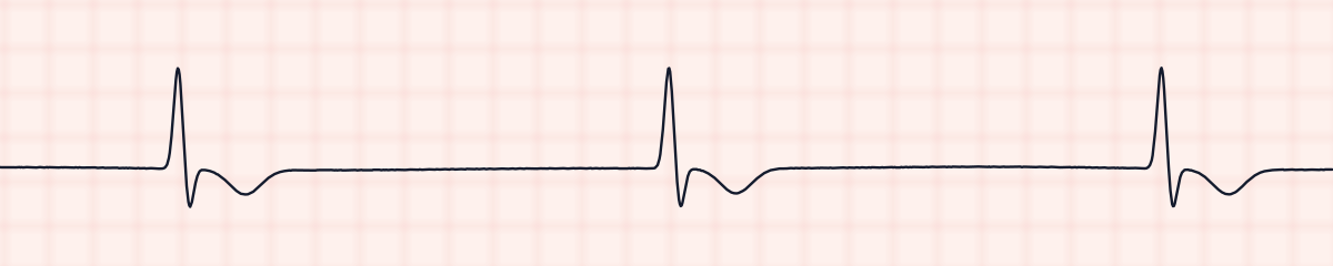
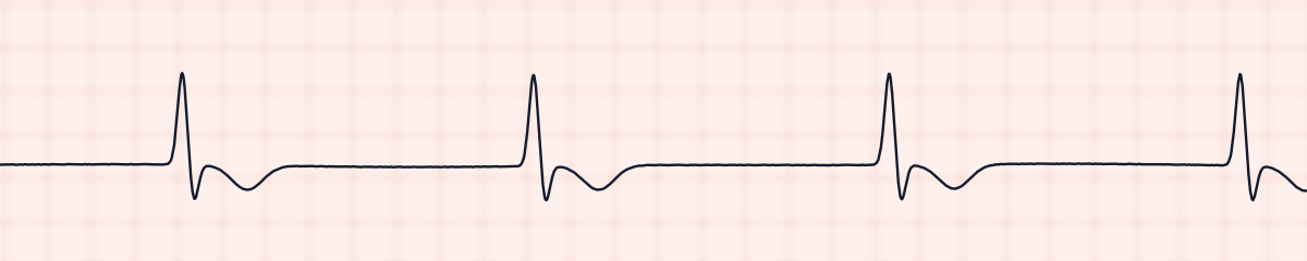
More idioventricular rhythm strips: no two are the same
A rhythm is a set of rules, not a picture. These three strips all meet the rules for idioventricular rhythm, with different numbers.
What idioventricular rhythm gets mistaken for
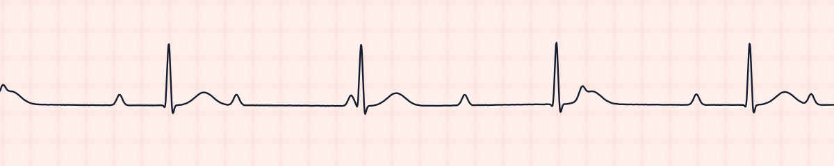
Idioventricular rhythm vs. third-degree (complete) AV block
Complete heart block with a ventricular escape has the same slow wide QRS, plus regular P waves that march through at their own faster rate. No P waves at all means idioventricular rhythm.
Idioventricular rhythm vs. ventricular tachycardia
The complexes have the same wide shape. The difference is rate: idioventricular rhythm is 20 to 40, accelerated idioventricular rhythm 40 to 100, and ventricular tachycardia is over 100.
Practice: idioventricular rhythm or a look-alike?
Five strips. Some show idioventricular rhythm; the others are the rhythms most often confused with it.
Strip 1 of 5
Lead II · 25 mm/s · 6 s
Causes of idioventricular rhythm
- Myocardial infarction and severe ischemia
- The minutes after blood flow is restored to a blocked coronary artery (typically AIVR)
- Drug toxicity, including digoxin
- Severe electrolyte and acid-base disturbances
- A dying heart: it is often seen late in a cardiac arrest
Why it matters
This is the heart’s last backup. At 20 to 40 beats per minute cardiac output is usually too low, and the patient may be hypotensive, unresponsive or pulseless. An idioventricular rhythm with no pulse is pulseless electrical activity. The accelerated form after reperfusion is different: it is usually brief, well tolerated and left alone.
What is done for idioventricular rhythm
- Check for a pulse and assess perfusion immediately.
- No pulse: start CPR and treat as PEA.
- With a pulse and symptoms: atropine may be tried, with pacing and dopamine or epinephrine infusions as support.
- Do not give drugs that suppress ventricular beats, such as lidocaine or amiodarone. This rhythm is the only thing keeping the ventricles beating, and suppressing it can cause asystole.
- Look for and treat the cause.
Study summary, not a protocol. Follow current American Heart Association guidelines and your facility’s policies. See the medical disclaimer.
Common mistakes with idioventricular rhythm
- Calling it ventricular tachycardia because the QRS is wide. Count the rate first.
- Not searching for P waves, and missing a complete heart block.
- Reaching for an antiarrhythmic because the complexes “look ventricular.” Escape rhythms are supported, not suppressed.
Questions about idioventricular rhythm
Is an idioventricular rhythm dangerous?
At 20 to 40 beats per minute it usually is. A rate that slow often cannot hold up the blood pressure, and it means the faster pacemakers above the ventricles have stopped working. The first thing to do is feel for a pulse: an idioventricular rhythm without one is a cardiac arrest. The accelerated form, AIVR, is the exception, and is usually short and harmless.
How is an idioventricular rhythm treated?
By supporting it and fixing what caused it, never by suppressing it. With no pulse it is treated as PEA, with CPR. With a pulse and low blood pressure, atropine may be tried, and pacing or an epinephrine or dopamine infusion can hold the rate up while the cause is found. Follow your facility’s protocols.
What is the rate of an idioventricular rhythm?
20 to 40 beats per minute. That is the built-in rate of pacemaker cells in the ventricles.
What is accelerated idioventricular rhythm (AIVR)?
The same wide-QRS ventricular rhythm at a faster rate, about 40 to 100 per minute. It is common for a short time after a blocked coronary artery is reopened and usually needs no treatment.
Why should you not give lidocaine or amiodarone for an idioventricular rhythm?
Those drugs suppress impulses that start in the ventricles. In an escape rhythm the ventricles are the only pacemaker still working, so suppressing them can leave the patient with no rhythm at all.
Does an idioventricular rhythm have P waves?
No. If regular P waves are present and unrelated to the wide QRS complexes, the rhythm is complete heart block with a ventricular escape rhythm.