Ventricular rhythms
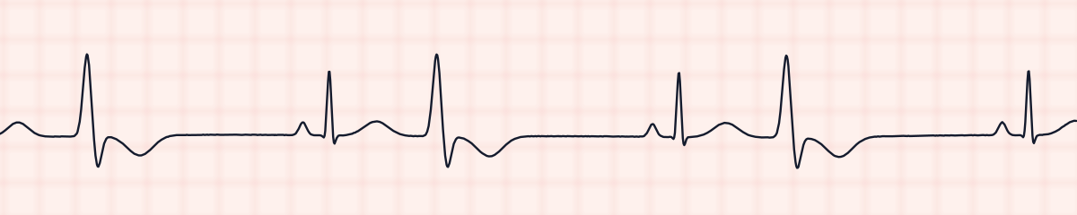
Multifocal PVCs on an EKG strip
Multifocal PVCs are premature ventricular contractions of two or more different shapes on the same strip. Each is early, wide and without a P wave, but they do not match: one may point up and another down, or they may differ in width and height. The different shapes show that they arise from different sites in the ventricles. They are also called multiform PVCs.
Criteria from the American Heart Association guidelines and published ECG standards · Every strip checked by measurement · Sources and method · Updated November 15, 2026
- Rate
- Underlying rhythm
- PR interval
- None on the PVCs
- QRS
- Wide PVCs of different shapes
How to identify multifocal PVCs in five steps
Ask the same five questions of every strip. The right-hand column is what each one finds on the strip above.
1Rate
That of the underlying rhythm.
~70 bpm
2Rhythm
Regular, interrupted by early beats and the pauses after them.
Regular, with early beats
3P waves
Normal on the sinus beats; none before any PVC.
None on the early beats
4PR interval
Normal on the sinus beats.
0.14 s on sinus beats
5QRS
Narrow on the sinus beats. The PVCs are wide and differ from each other in shape.
0.08 s; early beats 0.16 s, two shapes
What is happening in the heart
The shape of a ventricular beat depends on where in the ventricle it starts, because that sets the direction in which the impulse spreads. PVCs from one site all look the same. When more than one site is firing, the PVCs take on more than one shape, and often the time from the previous normal beat differs too.
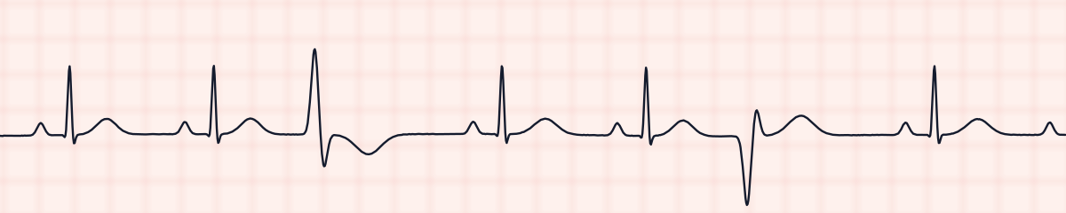
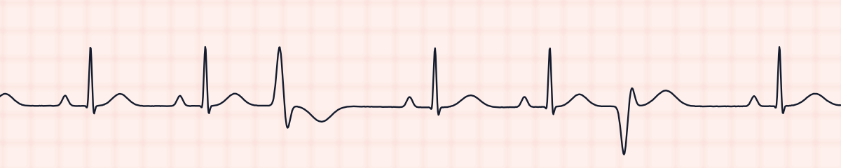
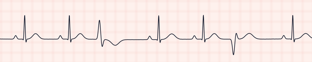
More multifocal PVC strips: no two are the same
A rhythm is a set of rules, not a picture. These three strips all meet the rules for multifocal PVCs, with different numbers.
What multifocal PVCs get mistaken for
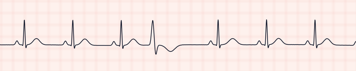
Multifocal PVCs vs. premature ventricular contractions (PVCs)
Unifocal PVCs are copies of each other. Lay one PVC over the next in your mind: if the direction or outline differs, they are multifocal.
Multifocal PVCs vs. ventricular bigeminy
Bigeminy describes timing, a PVC after every normal beat. Multifocal describes shape. A strip can be both; name the shape whenever the PVCs differ.
Practice: multifocal PVCs or a look-alike?
Five strips. Some show multifocal PVCs; the others are the rhythms most often confused with it.
Strip 1 of 5
Lead II · 25 mm/s · 6 s
Causes of multifocal PVCs
- Hypoxia
- Electrolyte disturbance, particularly low potassium and magnesium
- Acute ischemia
- Digoxin toxicity
- Severe heart failure
- Acid-base imbalance
Why it matters
With unifocal PVCs a single small patch of muscle is misfiring. More than one irritable site means the whole ventricle is unsettled. Multifocal PVCs are considered more worrying than unifocal ones and are on the standard list of PVC warning signs, because they more often precede ventricular tachycardia or fibrillation.
What is done for multifocal PVCs
- Assess the patient, including oxygen saturation.
- Report multifocal PVCs; do not wait to see whether they persist.
- Expect electrolytes to be checked and corrected.
- Review medications, including digoxin.
- Monitor closely for couplets and runs.
Study summary, not a protocol. Follow current American Heart Association guidelines and your facility’s policies. See the medical disclaimer.
Common mistakes with multifocal PVCs
- Not comparing the PVCs with each other, and reporting them simply as “PVCs.”
- Mistaking a differently shaped PVC for artifact.
- Confusing the terms: unifocal and multifocal are about shape, bigeminy and trigeminy about pattern.
Questions about multifocal PVCs
What does multifocal mean?
It means the premature beats start from more than one focus, or site, in the ventricles. Each site produces its own shape of beat, so the PVCs on the strip do not match. When they all match, they are called unifocal.
Are multifocal PVCs serious?
They are treated as a warning sign. Several irritable sites suggest a more unstable ventricle, so multifocal PVCs are reported and a cause such as low oxygen or an electrolyte problem is sought.
How can you tell PVCs are multifocal on a strip?
Compare their shapes in the same lead. If one PVC is upright and another inverted, or their outlines clearly differ, they are multifocal.