Supraventricular rhythms
Supraventricular tachycardia (SVT) EKG strip
Supraventricular tachycardia (SVT) is a fast rhythm that starts above the ventricles. On an EKG strip it is narrow, perfectly regular and very fast, usually 150 to 250 beats per minute, and P waves cannot be seen because they are buried in the QRS or the T wave. It typically starts and stops abruptly.
Criteria from the American Heart Association guidelines and published ECG standards · Every strip checked by measurement · Sources and method · Updated November 15, 2026
- Rate
- 150–250
- PR interval
- Not measurable
- QRS
- < 0.12 s
How to identify SVT in five steps
Ask the same five questions of every strip. The right-hand column is what each one finds on the strip above.
1Rate
150 to 250 per minute. The rate does not drift; it holds one value.
211 bpm
2Rhythm
Perfectly regular.
Regular
3P waves
Usually not visible. They may show as a small notch at the end of the QRS.
Not visible
4PR interval
Not measurable.
Not measurable
5QRS
Narrower than 0.12 s.
0.07 s
What is happening in the heart
In the most common form, an electrical impulse gets caught in a short circuit in or near the AV node and keeps circling. Each lap sends one impulse down to the ventricles through the normal pathway, so the QRS is narrow, and one back up to the atria at almost the same moment, so the P wave is hidden. Because one circuit sets the pace, the rhythm is as regular as a metronome.
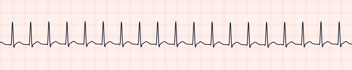
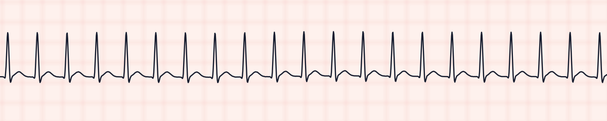
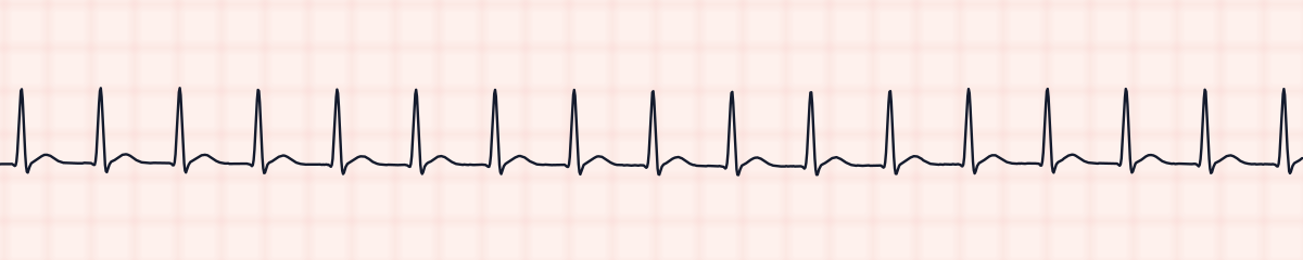
More SVT strips: no two are the same
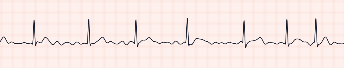
A rhythm is a set of rules, not a picture. These three strips all meet the rules for SVT, with different numbers.
What SVT gets mistaken for
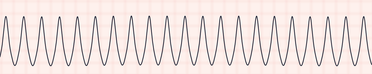
SVT vs. ventricular tachycardia
Both are fast and regular. Measure the QRS: narrower than 0.12 s points to SVT, 0.12 s or wider is treated as ventricular tachycardia until proven otherwise.
SVT vs. atrial fibrillation
Atrial fibrillation with a rapid response can look regular at a glance. Measure several R-R intervals. If they vary, it is not SVT.
SVT vs. normal sinus rhythm
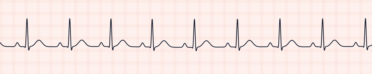
Sinus tachycardia has the shape of normal sinus rhythm at a faster rate: a visible P wave before each QRS, a rate that seldom passes 150 at rest, and a gradual speeding up and slowing down. SVT switches on and off.
Practice: SVT or a look-alike?
Five strips. Some show SVT; the others are the rhythms most often confused with it.
Strip 1 of 5
Lead II · 25 mm/s · 6 s
Causes of SVT
- An extra electrical pathway present from birth
- Caffeine, nicotine and other stimulants
- Stress and lack of sleep
- Alcohol
- Often no identifiable trigger, in an otherwise healthy heart
Why it matters
At these rates the ventricles have too little time to fill between beats, so cardiac output falls. Patients describe a pounding or fluttering chest, lightheadedness, shortness of breath or chest discomfort. Many tolerate it; some become hypotensive. It is usually not life-threatening in a healthy heart, but it does need to be stopped.
What is done for SVT
- Decide whether the patient is stable or unstable.
- Stable: vagal maneuvers first, such as a modified Valsalva.
- If that fails, adenosine by rapid IV push followed by a flush. Warn the patient: it causes a brief, unpleasant pause.
- Unstable (low blood pressure, altered mental status, chest pain, signs of shock): synchronized cardioversion.
- Run a continuous strip during adenosine; the rhythm as it breaks helps confirm the diagnosis.
Study summary, not a protocol. Follow current American Heart Association guidelines and your facility’s policies. See the medical disclaimer.
Common mistakes with SVT
- Calling any fast narrow rhythm SVT. Look for P waves and for how the rhythm started before ruling out sinus tachycardia.
- Treating sinus tachycardia as an arrhythmia. It is a response to something else, such as fever, pain, bleeding or dehydration, and the treatment is to fix the cause.
- Forgetting to measure the QRS, and missing a wide-complex tachycardia.
Questions about SVT
What heart rate is SVT?
Usually between 150 and 250 beats per minute. A narrow regular rhythm above about 150 at rest, with no visible P waves, should make you think of SVT.
How can you tell SVT from sinus tachycardia?
Sinus tachycardia has a P wave before every QRS, speeds up and slows down gradually, and rarely exceeds about 150 at rest. SVT starts and stops suddenly, holds a fixed rate, and its P waves are hidden.
Why does adenosine work for SVT?
Adenosine briefly blocks conduction through the AV node. Because the most common SVT circuits run through the node, interrupting it for a few seconds breaks the loop and lets the sinus node take over again.