Arrest rhythms
Ventricular fibrillation (V-fib) EKG strip
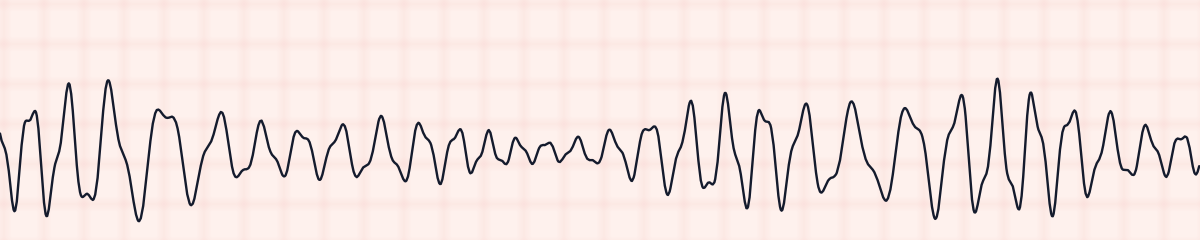
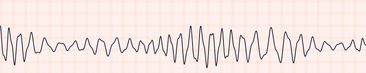
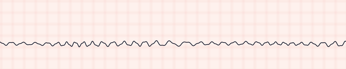
Ventricular fibrillation (VF or V-fib) is a cardiac arrest rhythm in which the ventricles quiver instead of pumping. On an EKG strip it is a chaotic, irregular wavy line with no P waves, no QRS complexes and no T waves. There is nothing that can be measured, and the patient has no pulse.
Criteria from the American Heart Association guidelines and published ECG standards · Every strip checked by measurement · Sources and method · Updated November 15, 2026
- Rate
- None
- PR interval
- None
- QRS
- None
How to identify ventricular fibrillation in five steps
Ask the same five questions of every strip. The right-hand column is what each one finds on the strip above.
1Rate
Cannot be counted.
None
2Rhythm
Chaotic. No two waves are alike in height, width or spacing.
Chaotic
3P waves
None.
None
4PR interval
None.
None
5QRS
None. There are only irregular undulations.
None
What is happening in the heart
Electrical activity in the ventricles breaks into many disorganized wavelets that fire at random. No coordinated contraction happens, so no blood is pumped. Early on the waves are large (coarse VF). As the heart muscle runs out of energy they shrink (fine VF) and eventually flatten into asystole, which is why the chance of a successful shock falls with every minute.
More ventricular fibrillation strips: no two are the same
A rhythm is a set of rules, not a picture. These three strips all meet the rules for ventricular fibrillation, with different numbers.
What ventricular fibrillation gets mistaken for
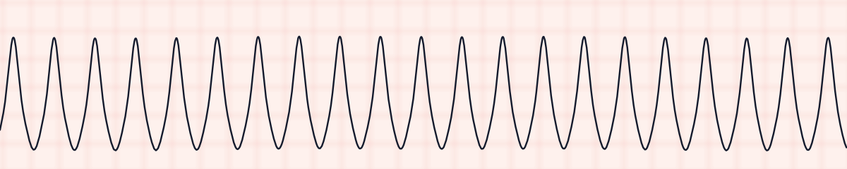
Ventricular fibrillation vs. ventricular tachycardia
Ventricular tachycardia has complexes of one repeating shape at an even spacing. In ventricular fibrillation nothing repeats.
Ventricular fibrillation vs. asystole
Fine ventricular fibrillation can be nearly flat. Turn up the gain and check a second lead: any persistent chaotic movement is VF and is shockable; a truly flat line is asystole and is not.
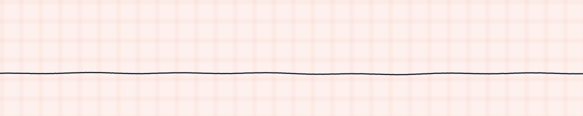
Practice: ventricular fibrillation or a look-alike?
Five strips. Some show ventricular fibrillation; the others are the rhythms most often confused with it.
Strip 1 of 5
Lead II · 25 mm/s · 6 s
Causes of ventricular fibrillation
- Myocardial infarction and ischemia, the most common cause
- Ventricular tachycardia that has not been treated
- Severe potassium or magnesium imbalance
- Electrical shock
- Drowning, hypothermia and severe hypoxia
- Drug toxicity and overdose
Why it matters
Ventricular fibrillation is cardiac arrest. There is no cardiac output, and brain injury begins within minutes. It is also the arrest rhythm with the best chance of survival, because it responds to defibrillation, but only if the shock is delivered quickly. This is one of the rhythms that competency exams require you to get right every time.
What is done for ventricular fibrillation
- Confirm the patient is unresponsive with no pulse, and call for help.
- Start high-quality CPR immediately.
- Defibrillate as soon as a defibrillator is available, then resume CPR at once.
- Epinephrine is given during the resuscitation, and amiodarone or lidocaine if VF persists after shocks.
- Look for reversible causes while CPR continues.
Study summary, not a protocol. Follow current American Heart Association guidelines and your facility’s policies. See the medical disclaimer.
Common mistakes with ventricular fibrillation
- Mistaking artifact for VF. A patient who is awake, talking or has a pulse is not in ventricular fibrillation: check the patient and the electrodes.
- Calling fine VF asystole and withholding a shock.
- Delaying the shock to do something else. Early defibrillation is the treatment.
Questions about ventricular fibrillation
What does V-fib look like on an EKG strip?
A chaotic, wavy line that never repeats. There are no P waves, no QRS complexes and no T waves, and the rate cannot be counted.
Is ventricular fibrillation shockable?
Yes. Ventricular fibrillation and pulseless ventricular tachycardia are the two shockable cardiac arrest rhythms. Asystole and PEA are not shockable.
What is the difference between coarse and fine V-fib?
Coarse VF has larger waves and usually means the arrest is recent. Fine VF has small waves, means the heart has been fibrillating longer, and is less likely to respond to a shock.
What is the difference between V-tach and V-fib?
Ventricular tachycardia is organized: wide complexes of the same shape at an even rate, sometimes with a pulse. Ventricular fibrillation is disorganized, has no complexes, and never has a pulse.