Sinus rhythms
EKG Artifact on a Rhythm Strip
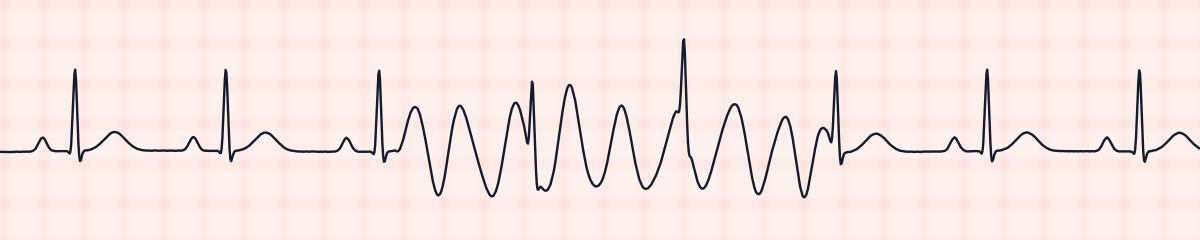
Artifact is anything on an EKG strip that the heart did not produce. Movement, muscle tremor, loose electrodes and electrical interference all leave marks on the trace, and some of those marks look like an arrhythmia. The kind that matters most is a wild, wide, fast stretch that resembles ventricular tachycardia or ventricular fibrillation. It is artifact, and not a rhythm, when the patient’s ordinary QRS complexes can still be found inside it, arriving on time.
Criteria from the American Heart Association guidelines and published ECG standards · Every strip checked by measurement · Sources and method · Updated November 15, 2026
- Rate
- That of the rhythm underneath
- PR interval
- 0.12–0.20 s where it can be seen
- QRS
- < 0.12 s, on time through the artifact
How to identify artifact in five steps
Ask the same five questions of every strip. The right-hand column is what each one finds on the strip above.
1Rate
Take it from the clean parts of the strip, or from the QRS spikes found inside the artifact. Disregard the number on the monitor: it is counting the artifact.
88 bpm
2Rhythm
Set calipers to the R-R interval on a clean stretch and step them into the artifact. If a narrow spike turns up at each step, the rhythm never changed.
Regular: the QRS complexes keep time through the artifact
3P waves
Present before each QRS wherever the baseline is clear. Inside the artifact they cannot be made out.
1 per QRS where the baseline is clear
4PR interval
Measured on the clean beats only.
0.13 s
5QRS
Narrow, and the same shape inside the artifact as outside it. The complexes are sharper than the artifact’s own waves, which is how they are picked out.
0.08 s
What is happening in the heart
A monitor amplifies tiny voltages picked up at the skin, and it amplifies whatever else changes the voltage at an electrode along with them. An arm that moves, a chest that shivers, a hand brushing teeth or patting a back, a cable that swings, an electrode that has dried out or lifted at one edge: each adds waves of its own to the heart’s. The heart is not affected. It goes on firing at its own rate, so its QRS complexes are still in the signal, sharp and evenly spaced, with the artifact drawn over and between them. The strips on this page show the kind most often mistaken for a ventricular rhythm: movement artifact over a sinus rhythm.
More artifact strips: no two are the same
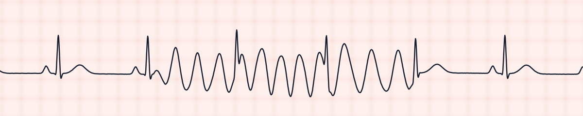
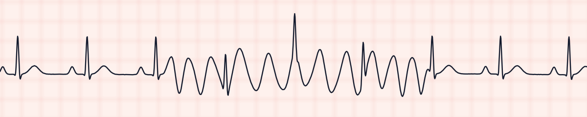
A rhythm is a set of rules, not a picture. These three strips all meet the rules for artifact, with different numbers.
What artifact gets mistaken for
Artifact vs. ventricular tachycardia
Ventricular tachycardia replaces the normal beats: every complex is wide and no narrow QRS shows among them. In artifact the narrow complexes run on through the wide waves at their earlier interval. And the patient looks well.
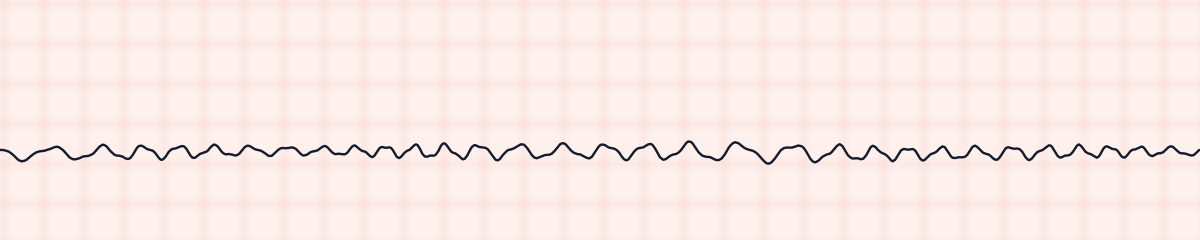
Artifact vs. ventricular fibrillation
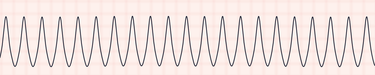
Ventricular fibrillation has no QRS complexes at all, and the patient is unresponsive with no pulse. Someone who is talking to you is not in V-fib, whatever the screen shows.
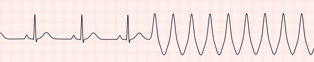
Artifact vs. sinus rhythm converting to ventricular tachycardia
When a sinus rhythm truly converts to V-tach, the narrow beats end at the first wide one. Under artifact they continue, and the clean rhythm that reappears afterwards is exactly in step with where it left off.
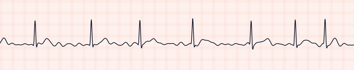
Artifact vs. atrial fibrillation
A fine muscle tremor makes the baseline fuzzy and can hide the P waves, which suggests atrial fibrillation. Check the R-R intervals: under a tremor they stay regular, and in fibrillation they never repeat.
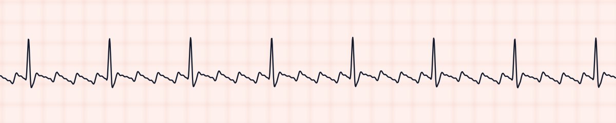
Artifact vs. atrial flutter
Even electrical interference, or a rhythmic tremor, can pass for flutter waves. True flutter waves keep a fixed relation to the QRS complexes and are still there when the patient lies still and the electrodes are changed.
Artifact vs. asystole
A lead that has come off draws a flat line. Asystole is named only in a patient with no pulse, once the leads and a second monitoring lead have been checked.
Practice: artifact or a look-alike?
Five strips. Some show artifact; the others are the rhythms most often confused with it.
Strip 1 of 5
Lead II · 25 mm/s · 6 s
Causes of artifact
- Patient movement: turning over, coughing, brushing teeth, combing hair, scratching
- Shivering, a tremor such as that of Parkinson’s disease, or tense muscles
- Chest physiotherapy, CPR and other rhythmic handling of the patient
- Electrodes that are dry, loose, or stuck over bone, hair or damp skin
- A broken lead wire or a loose connection
- Electrical interference from nearby equipment (60-cycle interference), which thickens the baseline with fine, perfectly even waves
- Breathing, which makes the whole trace rise and fall slowly (a wandering baseline)
Why it matters
Artifact is taken for a lethal rhythm often enough that patients have received unnecessary drugs, shocks and invasive procedures because of it. The opposite error happens too: a real arrhythmia is dismissed as artifact, or so many false alarms sound that staff stop answering them. Both are avoided in the same way, by looking at the patient first and then stepping the QRS complexes out across the strip. Rhythm exams include artifact for exactly this reason.
What is done for artifact
- Look at the patient before anything else. Awake, talking and with a pulse rules out ventricular fibrillation.
- Feel a pulse and compare its rate with the clean part of the strip.
- Find what is moving: the patient, a cable, a hand on the chest. Ask the patient to keep still and watch the trace settle.
- Check the electrodes. Replace any that are dry or loose, clean and dry the skin, clip hair if necessary, and place them over muscle and away from bones and joints.
- Check the lead wires and their connections, and move other electrical equipment away from the cables when the baseline is thick with fine, even waves.
- If any doubt remains that the rhythm is real, treat the patient as their condition requires and get another view: a second lead or a 12-lead ECG.
Study summary, not a protocol. Follow current American Heart Association guidelines and your facility’s policies. See the medical disclaimer.
Common mistakes with artifact
- Reading the monitor before reading the patient.
- Believing the displayed heart rate. The monitor counts the waves of the artifact as beats.
- Not stepping the R-R interval through the disturbed stretch.
- Silencing an alarm as “only artifact” without looking at either the strip or the patient.
- Leaving the cause in place. Artifact that is not corrected sets off the next false alarm as well.
Questions about artifact
What is artifact on an EKG?
Any deflection on the trace that does not come from the heart’s electrical activity. The usual sources are patient movement, muscle tremor, poor electrode contact and electrical interference.
How do you tell artifact from V-tach?
Measure the R-R interval on a clean part of the strip and step it through the suspicious part. With artifact, a narrow QRS spike appears at every step, on time, and the normal rhythm picks up in step afterwards. With V-tach the normal complexes are gone. Then check the patient: a pulse and a normal blood pressure go with artifact.
What are the types of EKG artifact?
Four are usually taught. Movement (motion) artifact gives large, irregular waves. Muscle tremor, also called somatic artifact, gives a fine, fuzzy, uneven baseline. 60-cycle (AC) interference gives a thick baseline of fine, perfectly even waves. A wandering baseline is the whole trace drifting up and down, usually with breathing. A loose or disconnected lead adds a fifth picture: a line that is flat or jumps about wildly.
What causes 60-cycle interference?
Alternating current in nearby equipment, such as infusion pumps, electric beds and other mains-powered devices, picked up through poorly attached electrodes or damaged cables. Better electrode contact and moving the cables away from the equipment usually clear it.
How do you fix a wandering baseline?
Make sure the electrodes are firmly stuck to clean, dry skin and that the cables are not tugging on them. Placing the electrodes where the chest wall moves least with breathing also helps.